Bos M P, Hogan D, Belland R J
Laboratory of Microbial Structure and Function, Rocky Mountain Laboratories, National Institute of Allergy and Infectious Diseases, Hamilton, Montana 59840-2999, USA.
J Exp Med. 1999 Aug 2;190(3):331-40. doi: 10.1084/jem.190.3.331.
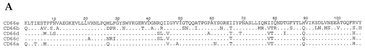
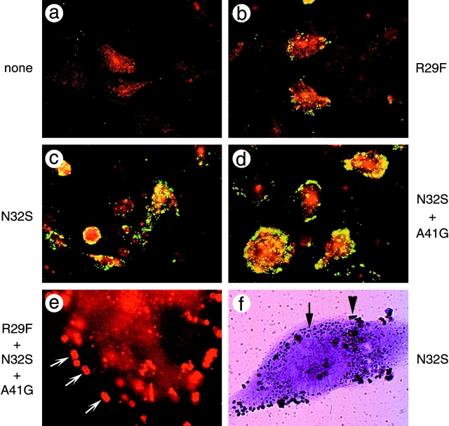
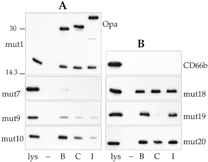
The immunoglobulin-like family of CD66 antigens, present on human neutrophils and epithelial cells, are used as receptors for adhesins expressed by the pathogenic Neisseriae. N. gonorrhoeae strain MS11 can express 11 isoforms of these adhesins, called opacity-related (Opa) proteins. Each MS11 Opa protein recognizes a distinct spectrum of CD66 receptors. CD66-Opa binding is mediated by the NH(2)-terminal domain of the receptor and occurs through protein-protein interactions. In this report, we have investigated the molecular basis for the binding between the CD66 and Opa protein families by mapping amino acids in CD66 receptors that determine Opa protein binding. We performed homologue scanning mutagenesis between CD66e, which binds multiple Opa variants, and CD66b, which binds none, and tested both loss-of-function by CD66e and gain-of-function by CD66b in solution assays and in assays involving full-length receptors expressed by epithelial cells. We found that three residues in the CD66e N-domain are required for maximal Opa protein receptor activity. Opa proteins that recognize the same spectrum of native CD66 molecules showed differential binding of receptors with submaximal activity, indicating that the binding characteristics of these Opa proteins are actually slightly different. These data provide a first step toward resolving the structural requirements for Opa-CD66 interaction.
存在于人类中性粒细胞和上皮细胞上的CD66抗原免疫球蛋白样家族,被用作致病性奈瑟菌表达的黏附素的受体。淋病奈瑟菌MS11菌株可表达11种这些黏附素的异构体,称为不透明相关(Opa)蛋白。每种MS11 Opa蛋白识别不同的CD66受体谱。CD66与Opa的结合由受体的NH(2)末端结构域介导,并通过蛋白质-蛋白质相互作用发生。在本报告中,我们通过绘制CD66受体中决定Opa蛋白结合的氨基酸,研究了CD66和Opa蛋白家族之间结合的分子基础。我们在结合多种Opa变体的CD66e和不结合任何Opa变体的CD66b之间进行了同源扫描诱变,并在溶液分析和涉及上皮细胞表达的全长受体的分析中测试了CD66e的功能丧失和CD66b的功能获得。我们发现CD66e N结构域中的三个残基是最大Opa蛋白受体活性所必需的。识别相同天然CD66分子谱的Opa蛋白显示出与次最大活性受体的差异结合,表明这些Opa蛋白的结合特性实际上略有不同。这些数据为解决Opa-CD66相互作用的结构要求迈出了第一步。