Correia I, Chu D, Chou Y H, Goldman R D, Matsudaira P
Whitehead Institute for Biomedical Research, Massachusetts Institute of Technology, Cambridge, Massachusetts 02142, USA.
J Cell Biol. 1999 Aug 23;146(4):831-42. doi: 10.1083/jcb.146.4.831.
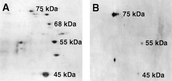
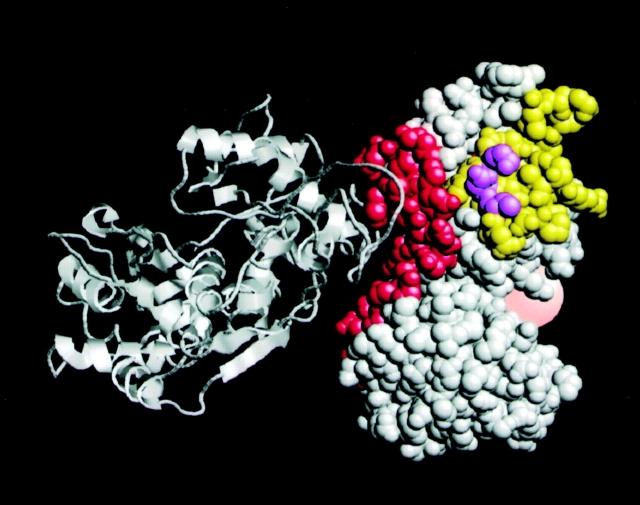
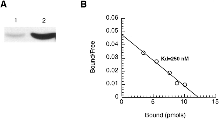
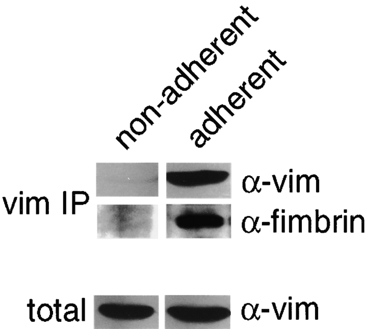
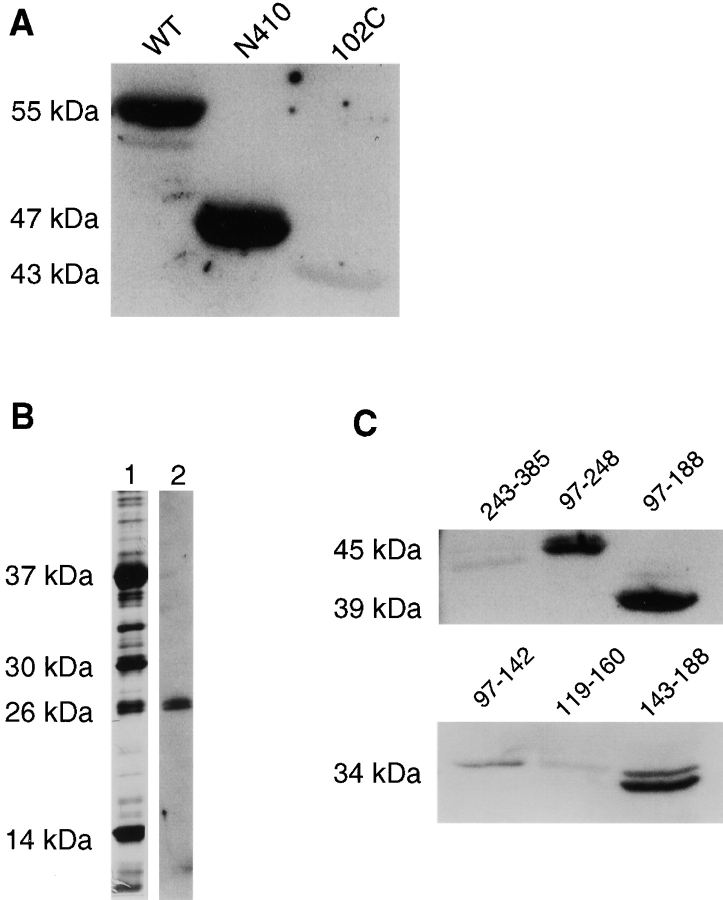
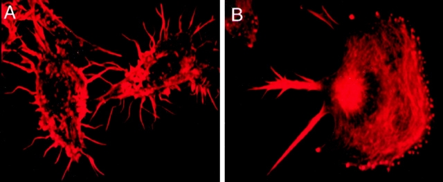
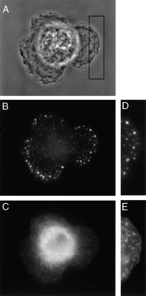
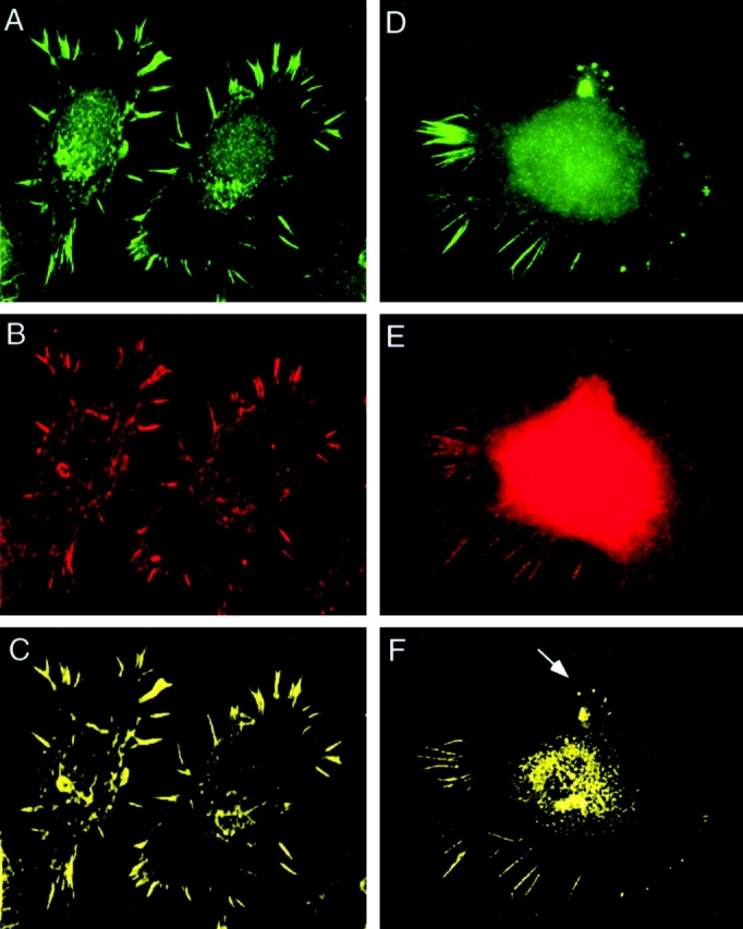
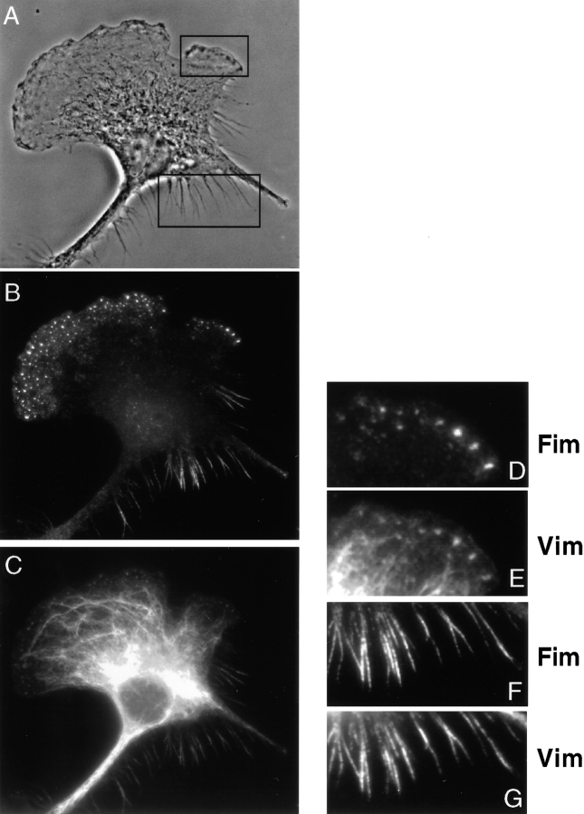
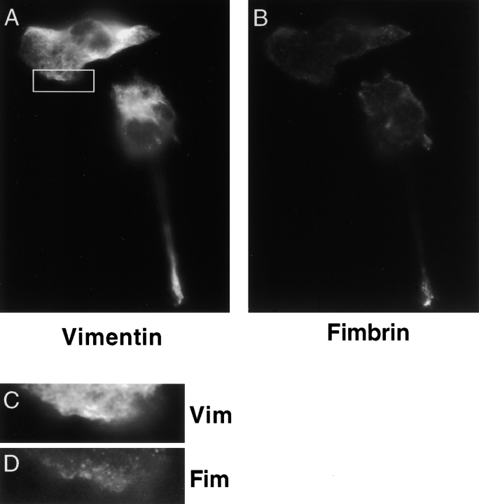
Cells adhere to the substratum through specialized structures that are linked to the actin cytoskeleton. Recent studies report that adhesion also involves the intermediate filament (IF) and microtubule cytoskeletons, although their mechanisms of interaction are unknown. Here we report evidence for a novel adhesion-dependent interaction between components of the actin and IF cytoskeletons. In biochemical fractionation experiments, fimbrin and vimentin coprecipitate from detergent extracts of macrophages using vimentin- or fimbrin-specific antisera. Fluorescence microscopy confirms the biochemical association. Both proteins colocalized to podosomes in the earliest stages of cell adhesion and spreading. The complex is also found in filopodia and retraction fibers. After detergent extraction, fimbrin and vimentin staining of podosomes, filopodia, and retraction fibers are lost, confirming that the complex is localized to these structures. A 1:4 stoichiometry of fimbrin binding to vimentin and a low percentage (1%) of the extracted vimentin suggest that fimbrin interacts with a vimentin subunit. A fimbrin-binding site was identified in the NH(2)-terminal domain of vimentin and the vimentin binding site at residues 143-188 in the CH1 domain of fimbrin. Based on these observations, we propose that a fimbrin-vimentin complex may be involved in directing the assembly of the vimentin cytoskeleton at cell adhesion sites.
细胞通过与肌动蛋白细胞骨架相连的特殊结构附着于基质。最近的研究报道,黏附还涉及中间丝(IF)和微管细胞骨架,尽管它们的相互作用机制尚不清楚。在此,我们报告肌动蛋白和IF细胞骨架成分之间存在一种新的黏附依赖性相互作用的证据。在生化分级分离实验中,使用波形蛋白或丝束蛋白特异性抗血清,丝束蛋白和波形蛋白从巨噬细胞的去污剂提取物中共沉淀。荧光显微镜证实了这种生化关联。在细胞黏附与铺展的最早阶段,两种蛋白质都共定位于足体。在丝状伪足和收缩纤维中也发现了这种复合物。去污剂提取后,足体、丝状伪足和收缩纤维的丝束蛋白和波形蛋白染色消失,证实该复合物定位于这些结构。丝束蛋白与波形蛋白的结合化学计量比为1:4,且提取的波形蛋白中有低比例(1%)与丝束蛋白结合,这表明丝束蛋白与波形蛋白的一个亚基相互作用。在波形蛋白的NH(2)末端结构域中鉴定出一个丝束蛋白结合位点,在丝束蛋白CH1结构域的143 - 188位残基处鉴定出波形蛋白结合位点。基于这些观察结果,我们提出丝束蛋白 - 波形蛋白复合物可能参与指导波形蛋白细胞骨架在细胞黏附位点的组装。