Holt S C, Gauther J J, Tipper D J
J Bacteriol. 1975 Jun;122(3):1322-38. doi: 10.1128/jb.122.3.1322-1338.1975.
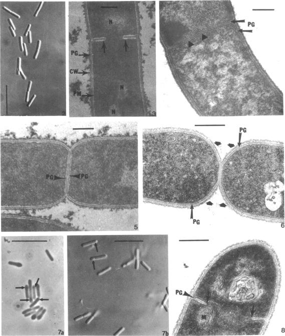
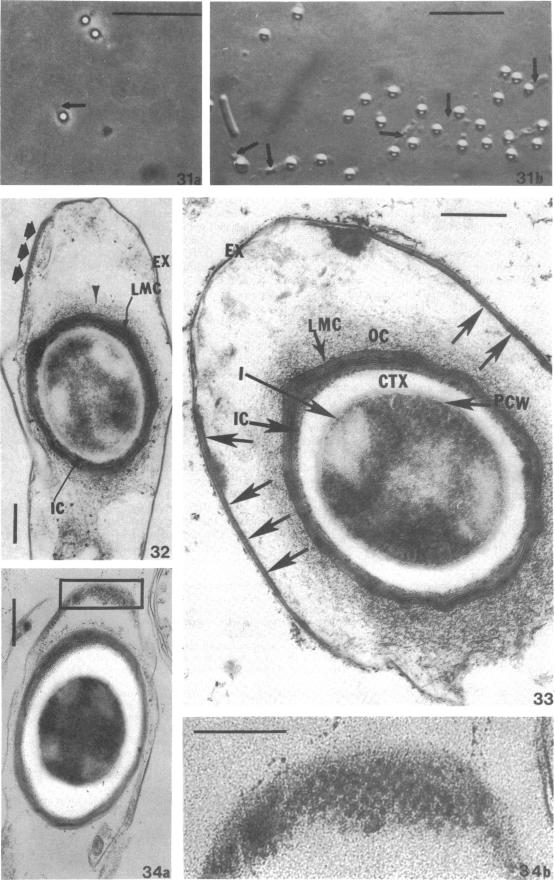
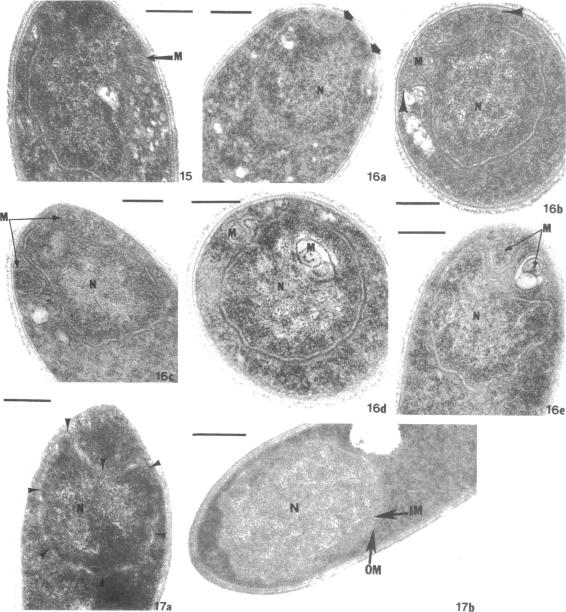
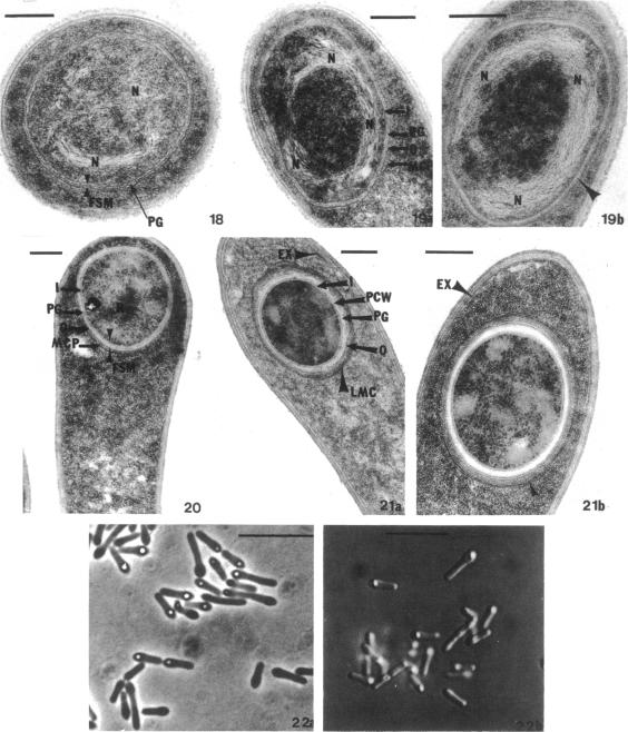
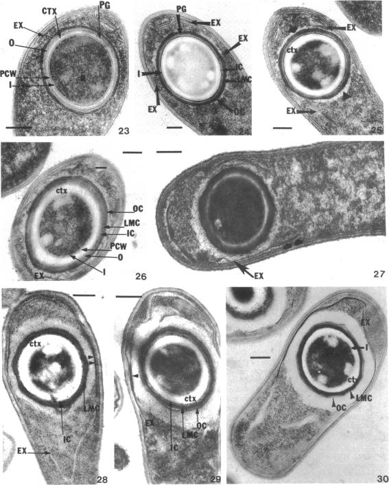
Spore septum formation in Bacillus sphaericus 9602 occurs 2 h after the end of exponential growth at one end of the vegetative cell, which retains a uniform diameter. The apparently rigid spore septum contains an inner cell wall layer which disappears when the sporulation septum "bulges" into the mother cell cytoplasm. This process occurs simultaneously with terminal swelling at the end of the cell containing the spore septum. It is suggested that the inner cell wall layer is peptidoglycan and that its dissolution and the terminal swelling are consequences of a localized autolysis. Engulfment of the forespore by membrane proliferation results in the production of a forespore surrounded by two flexible, closely apposed membranes. These membranes appear to become more rigid as a peptidoglycan-like layer appears between them, concomitant with the condensation of the forespore nucleoid into a crescent-shaped structure. After nuclear condensation, visible development of distinct cortex, primordial cell wall, and spore coat layers begin, and the forespore cytoplasm assumes an appearance similar to that of a refractile spore. The spore coats consist of an amorphous inner layer, a lamellar midlayer, and a structured outer layer. As cortex synthesis and spore coat assembly continue, exosporium development commences close to that portion of the mother cell plasma membrane which surrounds the forespore. The exosporium is lamellar and in tangential section is seen to have a hexagonal arrangement of subunits. The timing of these morphological events has the expected correlation with the appearance of unique enzyme activites required for cortex synthesis.
球形芽孢杆菌9602中的芽孢隔膜形成发生在指数生长结束后2小时,于营养细胞一端开始,该端细胞直径保持一致。明显坚硬的芽孢隔膜包含一层内部细胞壁层,当芽孢形成隔膜“鼓出”进入母细胞细胞质时,这层细胞壁层消失。此过程与含芽孢隔膜的细胞末端肿胀同时发生。据推测,内部细胞壁层是肽聚糖,其溶解和末端肿胀是局部自溶的结果。通过膜增殖对前芽孢的吞噬导致产生一个被两层灵活且紧密贴合的膜包围的前芽孢。随着它们之间出现一层类似肽聚糖的层,这些膜似乎变得更坚硬,同时前芽孢类核浓缩成新月形结构。核浓缩后,明显可见独特的皮层、原始细胞壁和芽孢衣层开始发育,前芽孢细胞质呈现出类似于折射性芽孢的外观。芽孢衣由无定形的内层、层状的中层和结构化的外层组成。随着皮层合成和芽孢衣组装的继续,芽孢外壁发育在靠近包围前芽孢的母细胞质膜部分开始。芽孢外壁是层状的,在切向切片中可见其亚基呈六边形排列。这些形态学事件的时间安排与皮层合成所需独特酶活性的出现具有预期的相关性。