Rabi Rebecca, Turnbull Lynne, Whitchurch Cynthia B, Awad Milena, Lyras Dena
Infection and Immunity Program, Monash Biomedicine Discovery Institute and Department of Microbiology, Monash University, Clayton, Victoria, Australia.
ithree institute, University of Technology Sydney, Ultimo, NSW, Australia.
mSphere. 2017 Oct 4;2(5). doi: 10.1128/mSphere.00343-17. eCollection 2017 Sep-Oct.
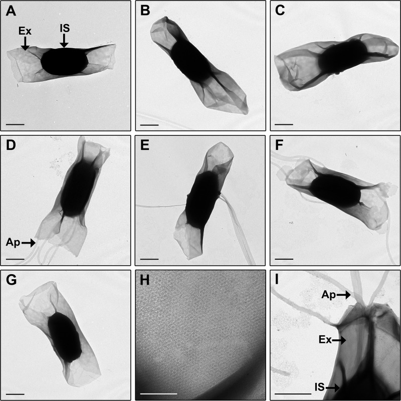
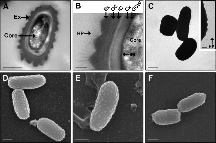
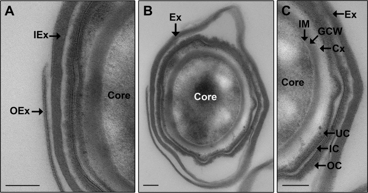
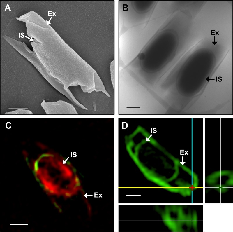
is an often-lethal bacterium causing human and animal disease. Crucial to the infectious cycle of is its ability to produce spores, which can germinate into toxin-producing vegetative bacteria under favorable conditions. However, structural details of the spore are lacking. Here, we used a range of electron microscopy techniques together with superresolution optical microscopy to characterize the spore morphology with an emphasis on the exosporium. The spore is made up of multiple layers with the exosporium presenting as a smooth balloon-like structure that is open at the spore poles. Focusing on the outer spore layers, we compared the morphologies of spores derived from different strains and determined that there is some variation between the spores, most notably with spores of some strains having tubular appendages. Since is a close relative of , their spores were compared by electron microscopy and their exosporia were found to be distinctly different from each other. This study therefore provides new structural details of the spore and offers insights into the physical structure of the exosporium across clostridial species. is a significant pathogen with mortality rates approaching 100%. It is the bacterial spore that is critical in initiating infection and disease. An understanding of spore structures as well as spore morphology across a range of strains may lead to a better understanding of infection and disease. However, the structural characteristics of the spores are limited. In this work, we have addressed this lack of detail and characterized the spore morphology. The use of traditional and advanced microscopy techniques has provided detailed new observations of spore structural features, which serve as a reference point for structural studies of spores from other bacterial species.
是一种常引发人类和动物疾病且往往致命的细菌。对其感染周期至关重要的是它产生孢子的能力,这些孢子在适宜条件下可萌发成产毒素的营养细菌。然而,关于该细菌孢子的结构细节尚不清楚。在此,我们使用了一系列电子显微镜技术以及超分辨率光学显微镜来表征该细菌孢子的形态,重点关注芽孢外壁。该细菌孢子由多层组成,芽孢外壁呈现为光滑的气球状结构,在孢子两极开口。聚焦于孢子外层,我们比较了不同菌株来源的该细菌孢子的形态,确定孢子之间存在一些差异,最显著的是一些菌株的孢子具有管状附属物。由于该细菌是[另一细菌名称]的近亲,通过电子显微镜对它们的孢子进行了比较,发现它们的芽孢外壁明显不同。因此,本研究提供了该细菌孢子的新结构细节,并深入了解了梭菌属各物种芽孢外壁的物理结构。该细菌是一种重要病原体,死亡率接近100%。正是细菌孢子在引发感染和疾病方面至关重要。了解一系列菌株的孢子结构以及孢子形态可能有助于更好地理解该细菌的感染和疾病。然而,该细菌孢子的结构特征有限。在这项工作中,我们解决了这一细节缺失问题,并表征了该细菌孢子的形态。传统和先进显微镜技术的使用提供了关于该细菌孢子结构特征的详细新观察结果,这些结果可作为其他细菌物种孢子结构研究的参考点。