Lee J Y, Qu-Petersen Z, Cao B, Kimura S, Jankowski R, Cummins J, Usas A, Gates C, Robbins P, Wernig A, Huard J
Growth and Development Laboratory, Department of Orthopaedic Surgery, Children's Hospital and University of Pittsburgh, Pittsburgh, Pennsylvania 15261, USA.
J Cell Biol. 2000 Sep 4;150(5):1085-100. doi: 10.1083/jcb.150.5.1085.
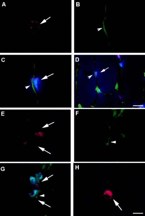
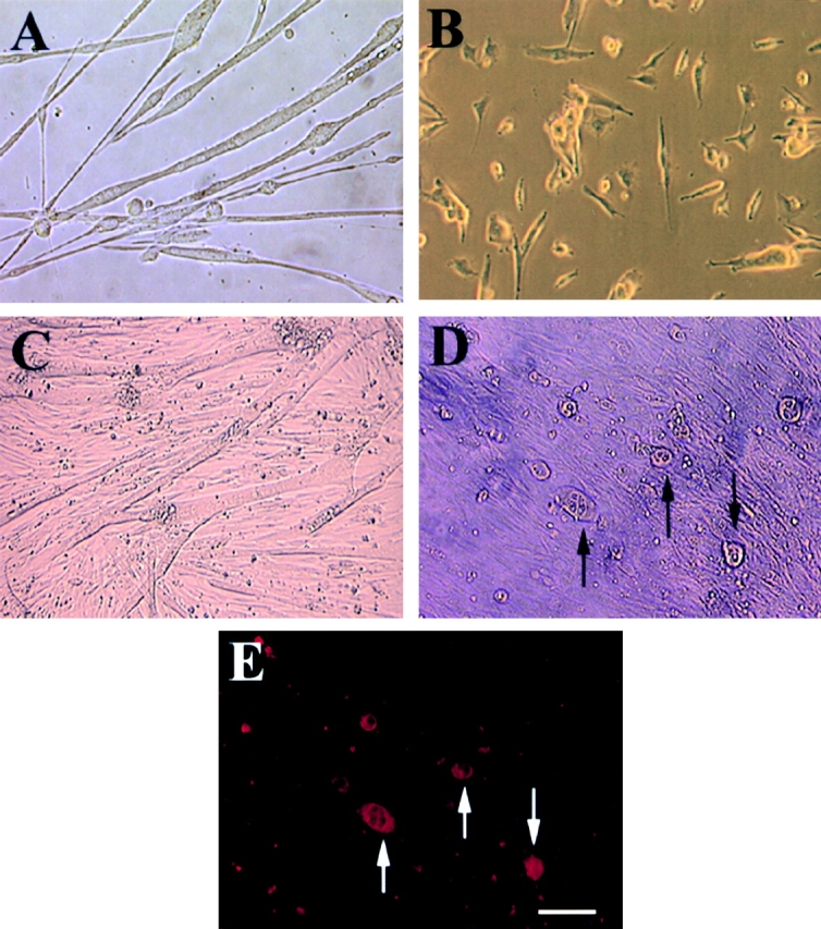
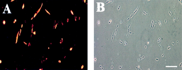
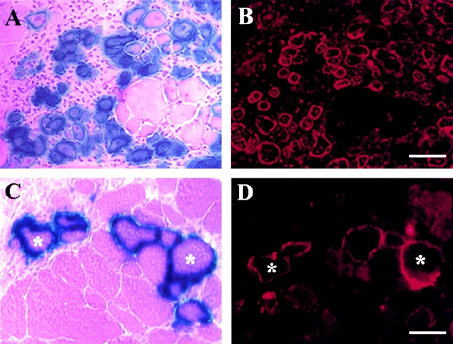
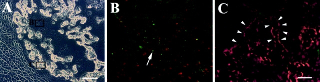
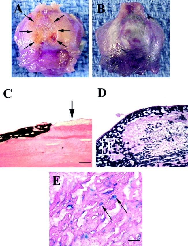
Several recent studies suggest the isolation of stem cells in skeletal muscle, but the functional properties of these muscle-derived stem cells is still unclear. In the present study, we report the purification of muscle-derived stem cells from the mdx mouse, an animal model for Duchenne muscular dystrophy. We show that enrichment of desmin(+) cells using the preplate technique from mouse primary muscle cell culture also enriches a cell population expressing CD34 and Bcl-2. The CD34(+) cells and Bcl-2(+) cells were found to reside within the basal lamina, where satellite cells are normally found. Clonal isolation and characterization from this CD34(+)Bcl-2(+) enriched population yielded a putative muscle-derived stem cell, mc13, that is capable of differentiating into both myogenic and osteogenic lineage in vitro and in vivo. The mc13 cells are c-kit and CD45 negative and express: desmin, c-met and MNF, three markers expressed in early myogenic progenitors; Flk-1, a mouse homologue of KDR recently identified in humans as a key marker in hematopoietic cells with stem cell-like characteristics; and Sca-1, a marker for both skeletal muscle and hematopoietic stem cells. Intramuscular, and more importantly, intravenous injection of mc13 cells result in muscle regeneration and partial restoration of dystrophin in mdx mice. Transplantation of mc13 cells engineered to secrete osteogenic protein differentiate in osteogenic lineage and accelerate healing of a skull defect in SCID mice. Taken together, these results suggest the isolation of a population of muscle-derived stem cells capable of improving both muscle regeneration and bone healing.
最近的几项研究表明骨骼肌中存在干细胞,但这些肌肉来源的干细胞的功能特性仍不清楚。在本研究中,我们报告了从mdx小鼠(杜兴氏肌营养不良症的动物模型)中纯化肌肉来源的干细胞。我们发现,使用预铺板技术从小鼠原代肌肉细胞培养物中富集结蛋白(desmin)阳性细胞,也能富集表达CD34和Bcl-2的细胞群体。发现CD34阳性细胞和Bcl-2阳性细胞位于基膜内,而卫星细胞通常也存在于此。从这个富集的CD34阳性Bcl-2阳性群体中进行克隆分离和鉴定,得到了一种假定的肌肉来源的干细胞mc13,它能够在体外和体内分化为成肌和成骨谱系。mc13细胞c-kit和CD45呈阴性,表达:结蛋白、c-met和MNF,这是早期成肌祖细胞中表达的三种标志物;Flk-1,KDR的小鼠同源物,最近在人类中被鉴定为具有干细胞样特征的造血细胞的关键标志物;以及Sca-1,骨骼肌和造血干细胞的标志物。肌肉内注射,更重要的是静脉注射mc13细胞可导致mdx小鼠的肌肉再生和肌营养不良蛋白的部分恢复。经基因工程改造分泌成骨蛋白的mc13细胞移植后可分化为成骨谱系,并加速SCID小鼠颅骨缺损的愈合。综上所述,这些结果表明分离出了一群能够改善肌肉再生和骨愈合的肌肉来源的干细胞。