• 文献检索
  • 文档翻译
  • 深度研究
  • 学术资讯
  • Suppr Zotero 插件Zotero 插件
  • 邀请有礼
  • 套餐&价格
  • 历史记录
应用&插件
Suppr Zotero 插件Zotero 插件浏览器插件Mac 客户端Windows 客户端微信小程序
定价
高级版会员购买积分包购买API积分包
服务
文献检索文档翻译深度研究API 文档MCP 服务
关于我们
关于 Suppr公司介绍联系我们用户协议隐私条款
关注我们

Suppr 超能文献

核心技术专利:CN118964589B侵权必究
粤ICP备2023148730 号-1Suppr @ 2026

文献检索

告别复杂PubMed语法,用中文像聊天一样搜索,搜遍4000万医学文献。AI智能推荐,让科研检索更轻松。

立即免费搜索

文件翻译

保留排版,准确专业,支持PDF/Word/PPT等文件格式,支持 12+语言互译。

免费翻译文档

深度研究

AI帮你快速写综述,25分钟生成高质量综述,智能提取关键信息,辅助科研写作。

立即免费体验

正常及经实验处理的田鼠(Microtus agrestis)垂体前叶的超微结构。

The ultrastructure of the anterior pituitary gland of the vole, Microtus agrestis, in normal and experimentally manipulated animals.

作者信息

Charlton H M, Worth R W

出版信息

J Anat. 1975 Sep;120(Pt 1):69-79.

PMID:1102504
原文链接:https://pmc.ncbi.nlm.nih.gov/articles/PMC1231724/
Abstract

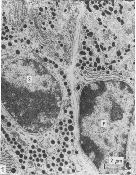
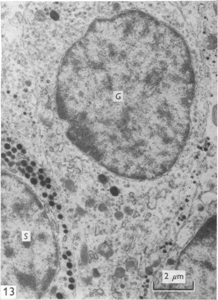
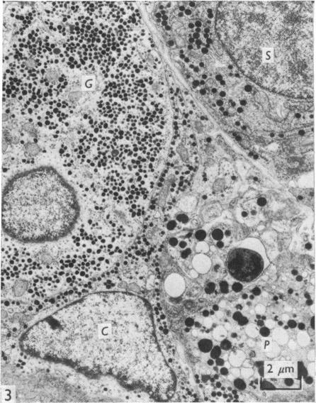
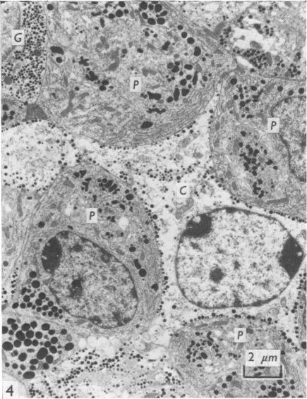
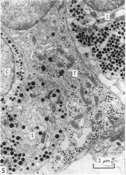
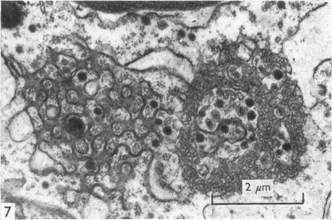
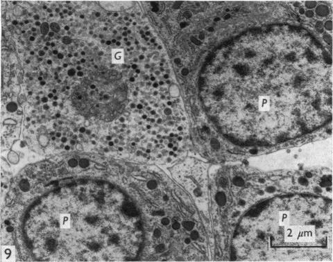
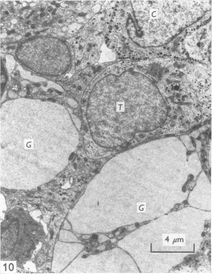
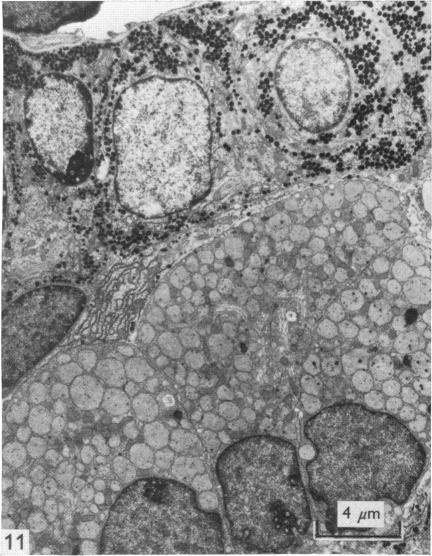
The ultrastructural appearance of the various types of cell present in the anterior pituitary of the vole has been described. There was a great measure of similarity between the cytological picture in this species and in the rat. Prolactotrophs contained the largest secretory granules, which were of variable shape; the granules of somatotrophs, whilst only slightly smaller than those of prolactotrophs, were invariably round, and of more uniform size; corticotrophs were represented by cells which were extremely angular, and whose secretory granules, besides being smaller than those of somatotrophs, were arrayed around the periphery of the cell below the plasma membrane; gonadotrophs contained granules of a similar size to those found in cortiocotrophs, but were found throughout the cytoplasm of the cells, whic were round to ovoid in shape; thyrotrophs contained the smallest granules of all, the shape of the cell itself bein angular...

摘要

已描述了田鼠垂体前叶中存在的各种类型细胞的超微结构外观。该物种的细胞学图像与大鼠的细胞学图像有很大程度的相似性。催乳素细胞含有最大的分泌颗粒,其形状各异;生长激素细胞的颗粒虽然仅比催乳素细胞的颗粒略小,但总是圆形的,且大小更均匀;促肾上腺皮质激素细胞由极具角状的细胞代表,其分泌颗粒除了比生长激素细胞的颗粒小之外,还排列在细胞膜下方细胞的周边;促性腺激素细胞含有与促肾上腺皮质激素细胞中发现的颗粒大小相似的颗粒,但存在于整个细胞的细胞质中,细胞呈圆形至椭圆形;促甲状腺激素细胞含有所有细胞中最小的颗粒,细胞本身呈角状……

https://cdn.ncbi.nlm.nih.gov/pmc/blobs/c509/1231724/de6345ebe080/janat00375-0081-d.jpg
https://cdn.ncbi.nlm.nih.gov/pmc/blobs/c509/1231724/8b9048579b88/janat00375-0078-a.jpg
https://cdn.ncbi.nlm.nih.gov/pmc/blobs/c509/1231724/4ff65b0a4728/janat00375-0078-b.jpg
https://cdn.ncbi.nlm.nih.gov/pmc/blobs/c509/1231724/f98d991b6df7/janat00375-0078-c.jpg
https://cdn.ncbi.nlm.nih.gov/pmc/blobs/c509/1231724/12ac42c02e1c/janat00375-0078-d.jpg
https://cdn.ncbi.nlm.nih.gov/pmc/blobs/c509/1231724/ebdd233ef422/janat00375-0080-a.jpg
https://cdn.ncbi.nlm.nih.gov/pmc/blobs/c509/1231724/4f87a555dd5e/janat00375-0080-b.jpg
https://cdn.ncbi.nlm.nih.gov/pmc/blobs/c509/1231724/0c816c8edf16/janat00375-0080-c.jpg
https://cdn.ncbi.nlm.nih.gov/pmc/blobs/c509/1231724/0781c8fbfe2f/janat00375-0080-d.jpg
https://cdn.ncbi.nlm.nih.gov/pmc/blobs/c509/1231724/7aa061705705/janat00375-0080-e.jpg
https://cdn.ncbi.nlm.nih.gov/pmc/blobs/c509/1231724/de1619e2b9fb/janat00375-0081-a.jpg
https://cdn.ncbi.nlm.nih.gov/pmc/blobs/c509/1231724/d7f39da6062b/janat00375-0081-b.jpg
https://cdn.ncbi.nlm.nih.gov/pmc/blobs/c509/1231724/6381cc00d275/janat00375-0081-c.jpg
https://cdn.ncbi.nlm.nih.gov/pmc/blobs/c509/1231724/de6345ebe080/janat00375-0081-d.jpg

相似文献

1
The ultrastructure of the anterior pituitary gland of the vole, Microtus agrestis, in normal and experimentally manipulated animals.正常及经实验处理的田鼠(Microtus agrestis)垂体前叶的超微结构。
J Anat. 1975 Sep;120(Pt 1):69-79.
2
Ultrastructure of the "non-pathologic" human pituitary gland.
Acta Pathol Jpn. 1976 Mar;26(2):191-203.
3
Recovery of rat prolactin cells following cessation of estrogen treatment.雌激素治疗停止后大鼠催乳素细胞的恢复
Anat Rec. 1976 May;185(1):31-48. doi: 10.1002/ar.1091850104.
4
Characterization of seasonal changes in prolactin and growth hormone cells in the hypophyses of white-tailed deer (Odocoileus borealis) by ultrastructural and immunocytochemical techniques.运用超微结构和免疫细胞化学技术对白尾鹿(北美白尾鹿)垂体中催乳素和生长激素细胞的季节性变化进行表征。
Am J Anat. 1981 Mar;160(3):277-84. doi: 10.1002/aja.1001600306.
5
SEASONAL CHANGES IN THE ADENOHYPOPHYSIS OF THE VOLE (MICROTUS AGRESTIS).
Gen Comp Endocrinol. 1964 Jun;4:243-52. doi: 10.1016/0016-6480(64)90018-8.
6
Ultrastructure of the cell types of the anterior hypophysis in a lizard. I. Corticotrophs.蜥蜴腺垂体细胞类型的超微结构。I.促肾上腺皮质激素细胞。
Arch Anat Microsc Morphol Exp. 1980;69(4):281-96.
7
The pars distalis (anterior pituitary) in the fetal sheep: an ultrastructural study.
J Dev Physiol. 1981 Oct;3(5):319-32.
8
Demonstration of the existence of a single morphological type of gonadotrophic cell in Ellobius lutescens (Microtinae) by an ultrastructural analysis of their development under various physiological and experimental conditions.
Cell Tissue Res. 1976 Mar 5;167(1):49-64. doi: 10.1007/BF00220159.
9
Ultrastructure of the fetal rat anterior pituitary gland as term and during prolonged gestation.足月及孕期延长时胎鼠垂体前叶的超微结构
Anat Rec. 1979 Jun;194(2):257-65. doi: 10.1002/ar.1091940207.
10
Distribution of gonadotrophins within the anterior pituitary cells of the musk shrew (Suncus murinus L.).促性腺激素在麝鼩(Suncus murinus L.)垂体前叶细胞内的分布。
Anat Rec. 1992 May;233(1):83-8. doi: 10.1002/ar.1092330111.

引用本文的文献

1
Fluctuation of CD9/SOX2-positive cell populations during the turnover of GH- and TSH-producing cells in the adult anterior pituitary gland.在成年垂体前叶 GH 和 TSH 产生细胞的更替过程中,CD9/SOX2 阳性细胞群体的波动。
J Reprod Dev. 2023 Dec 8;69(6):308-316. doi: 10.1262/jrd.2023-023. Epub 2023 Sep 30.

本文引用的文献

1
A SIMPLIFIED LEAD CITRATE STAIN FOR USE IN ELECTRON MICROSCOPY.一种用于电子显微镜的简化柠檬酸铅染色法。
J Cell Biol. 1965 May;25(2):407-8. doi: 10.1083/jcb.25.2.407.
2
SEASONAL CHANGES IN THE ADENOHYPOPHYSIS OF THE VOLE (MICROTUS AGRESTIS).
Gen Comp Endocrinol. 1964 Jun;4:243-52. doi: 10.1016/0016-6480(64)90018-8.
3
Electron microscope studies on the secretory cytology of the mouse anterior pituitary.小鼠垂体前叶分泌细胞学的电子显微镜研究
Endocrinology. 1962 Oct;71:618-28. doi: 10.1210/endo-71-4-618.
4
Electron microscopic studies of anterior pituitary glands from lactating and estrogen-treated rats.对哺乳期及经雌激素处理的大鼠垂体前叶进行的电子显微镜研究。
Endocrinology. 1961 Jul;69:81-90. doi: 10.1210/endo-69-1-81.
5
Adenohypophysis:prolactin.腺垂体:催乳素。
Annu Rev Physiol. 1966;28:57-88. doi: 10.1146/annurev.ph.28.030166.000421.
6
[The network of the folliculo-stellate cells and the follicles of the adenohypophysis in the rat (pars distalis)].[大鼠腺垂体远侧部的滤泡星状细胞网络与滤泡]
Z Zellforsch Mikrosk Anat. 1972;129(3):328-69.
7
Reviews of the progress of dairy science. Section A. Physiology. Hormonal control of lactogenesis.乳品科学进展综述。A部分。生理学。泌乳生成的激素调控。
J Dairy Res. 1971 Jun;38(2):237-64. doi: 10.1017/s0022029900019348.
8
The adenohypophysis of female rats after hypothalamic oestradiol implants: an electron microscopic study.下丘脑植入雌二醇后雌性大鼠的腺垂体:一项电子显微镜研究。
J Endocrinol. 1970 May;47(1):101-10. doi: 10.1677/joe.0.0470101.
9
Field and laboratory studies on the control of luteinizing hormone secretion and gonadal activity in the vole, Microtus agrestis.
J Reprod Fertil Suppl. 1973 Dec;19:89-99.
10
Adenohypophysis: ultrastructural cytochemistry. A review.腺垂体:超微结构细胞化学。综述。
J Histochem Cytochem. 1973 Oct;21(10):855-94. doi: 10.1177/21.10.855.