Elston D M
Department of Dermatology, Brooke Army Medical Center, Ft Sam, Houston, Texas 78234, USA.
BMC Microbiol. 2001;1:21. doi: 10.1186/1471-2180-1-21. Epub 2001 Sep 24.
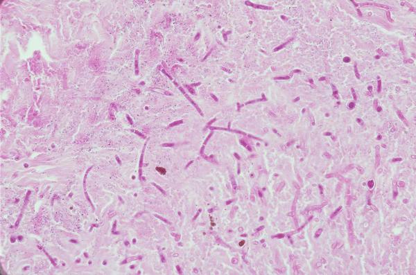
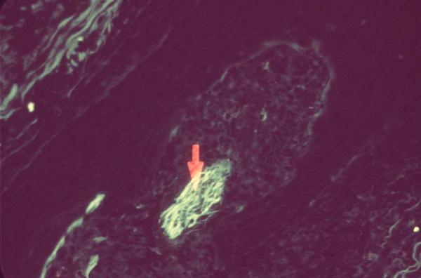
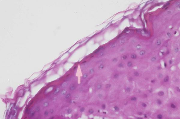
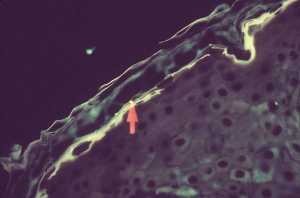
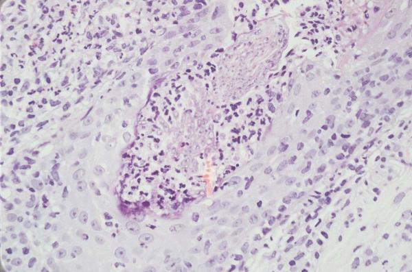
Fluorescence of many fungi is noted when H&E stained sections are examined under a fluorescent microscope. In theory, this phenomenon could aid in the diagnosis of cutaneous and disseminated fungal infections without the delay associated with special stains. Seventy-six cases of superficial and deep fungal infections and 3 cases of protothecosis were studied to determine the clinical usefulness of this technique.
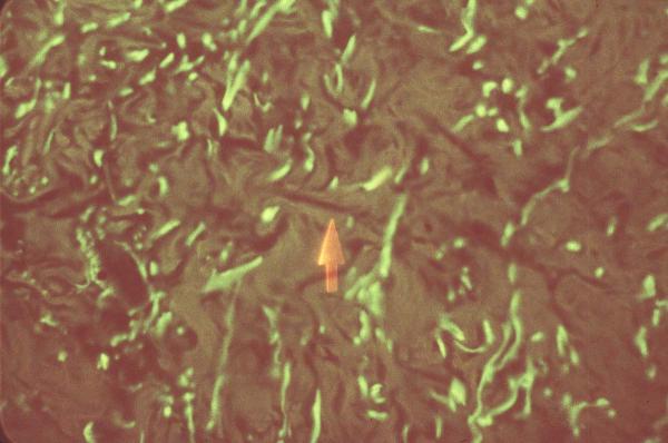
In most cases, fluorescence was noted, but was not intense. Fluorescence of fungi did not correlate with the age of the specimen. In most cases, organisms in H&E stained sections were more easily identified with routine light microscopy than with fluorescent microscopy.
This report suggests that in H&E stained skin specimens, fluorescent microscopy is of little benefit in the identification of fungal organisms.
当在荧光显微镜下检查苏木精-伊红(H&E)染色切片时,可观察到许多真菌发出荧光。理论上,这种现象有助于诊断皮肤和播散性真菌感染,而无需因特殊染色而延误诊断。本研究对76例浅表和深部真菌感染病例以及3例原藻病病例进行研究,以确定该技术的临床实用性。
在大多数病例中可观察到荧光,但并不强烈。真菌的荧光与标本的保存时间无关。在大多数情况下,H&E染色切片中的微生物通过常规光学显微镜比荧光显微镜更容易识别。
本报告表明,在H&E染色的皮肤标本中,荧光显微镜在识别真菌微生物方面益处不大。