Meunier J M, Trayanova N A, Gray R A
Department of Biomedical Engineering Tulane University, New Orleans, Louisiana 70118-5674, USA.
J Cardiovasc Electrophysiol. 2001 Oct;12(10):1176-84. doi: 10.1046/j.1540-8167.2001.01176.x.
Cardiac tissue can be entrained when subjected to sinusoidal stimuli, often responding with action potentials sustained for the duration of the stimulus. To investigate mechanisms responsible for both entrainment and extended action potential duration, computer simulations of a two-dimensional grid of cardiac cells subjected to sinusoidal extracellular stimulation were performed.
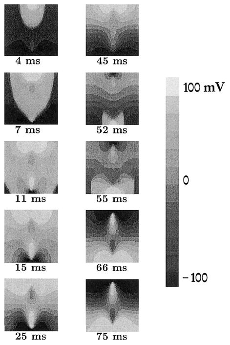
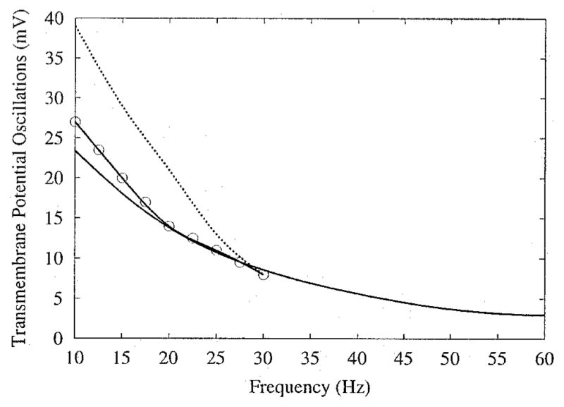
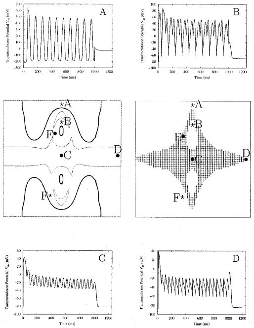
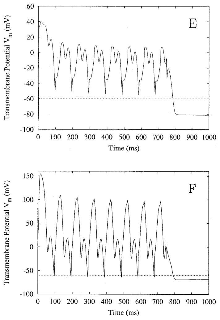
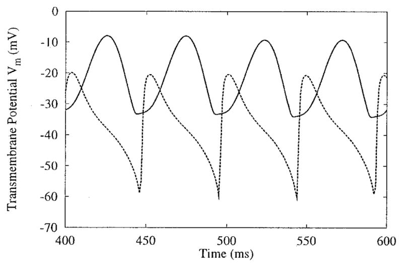
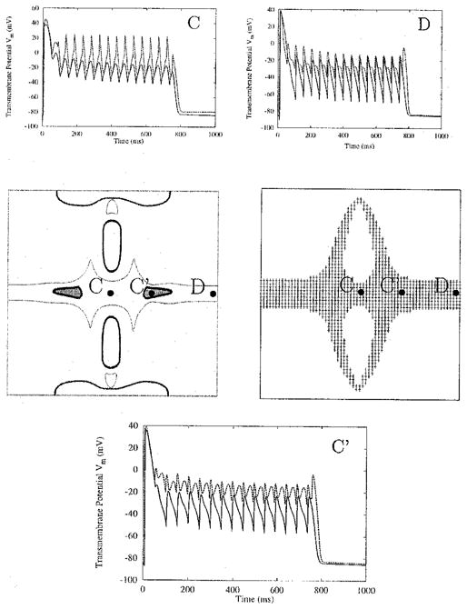
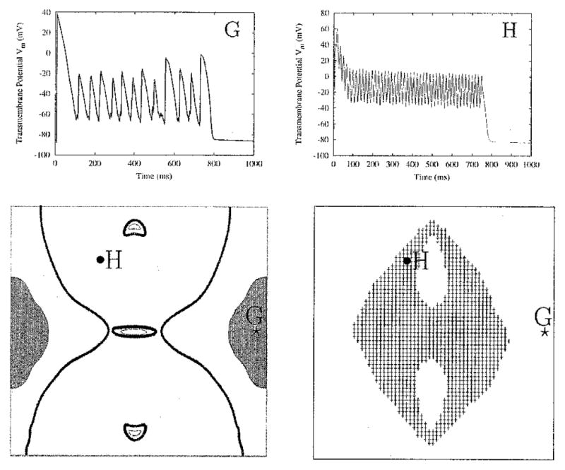
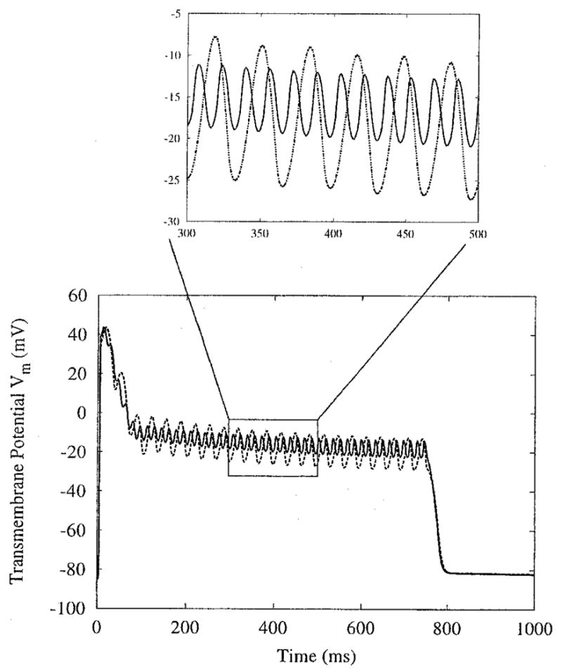
The tissue is represented as a bidomain with unequal anisotropy ratios. Cardiac membrane dynamics are governed by a modified Beeler-Reuter model. The stimulus, delivered by a bipolar electrode, has a duration of 750 to 1,000 msec, an amplitude range of 800 to 3,200 microA/cm, and a frequency range of 10 to 60 Hz. The applied stimuli create virtual electrode polarization (VEP) throughout the sheet. The simulations demonstrate that periodic extracellular stimulation results in entrainment of the tissue. This phase-locking of the membrane potential to the stimulus is dependent on the location in the sheet and the magnitude of the stimulus. Near the electrodes, the oscillations are 1:1 or 1:2 phase-locked; at the middle of the sheet, the oscillations are 1:2 or 1:4 phase-locked and occur on the extended plateau of an action potential. The 1:2 behavior near the electrodes is due to periodic change in the voltage gradient between VEP of opposite polarity; at the middle of the sheet, it is due to spread of electrotonic current following the collision of a propagating wave with refractory tissue.
The simulations suggest that formation of VEP in cardiac tissue subjected to periodic extracellular stimulation is of paramount importance to tissue entrainment and formation of an extended oscillatory action potential plateau.
心脏组织在受到正弦刺激时会被驱动,通常会产生持续整个刺激持续时间的动作电位。为了研究驱动和延长动作电位持续时间的机制,对受到正弦细胞外刺激的二维心脏细胞网格进行了计算机模拟。
组织被表示为具有不等各向异性比的双域。心脏膜动力学由改进的Beeler-Reuter模型控制。由双极电极施加的刺激持续时间为750至1000毫秒,幅度范围为800至3200微安/平方厘米,频率范围为10至60赫兹。施加的刺激在整个薄片中产生虚拟电极极化(VEP)。模拟表明,周期性细胞外刺激会导致组织被驱动。膜电位与刺激的这种锁相取决于薄片中的位置和刺激的大小。在电极附近,振荡为1:1或1:2锁相;在薄片中间,振荡为1:2或1:4锁相,并发生在动作电位的延长平台期。电极附近的1:2行为是由于相反极性的VEP之间电压梯度的周期性变化;在薄片中间,这是由于传播波与不应期组织碰撞后电紧张电流的传播。
模拟表明,在受到周期性细胞外刺激的心脏组织中VEP的形成对于组织驱动和延长振荡动作电位平台期的形成至关重要。