Hui Kwokyin, Lipkind Gregory, Fozzard Harry A, French Robert J
Department of Physiology and Biophysics, University of Calgary, 3330 Hospital Drive NW, Calgary, Alberta, Canada T2N 4N1.
J Gen Physiol. 2002 Jan;119(1):45-54. doi: 10.1085/jgp.119.1.45.
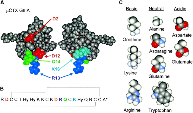
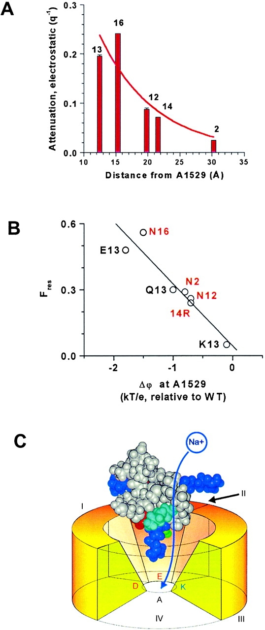
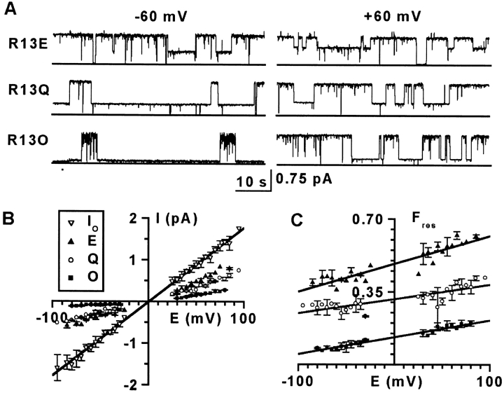
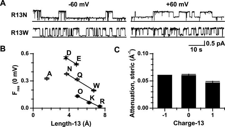
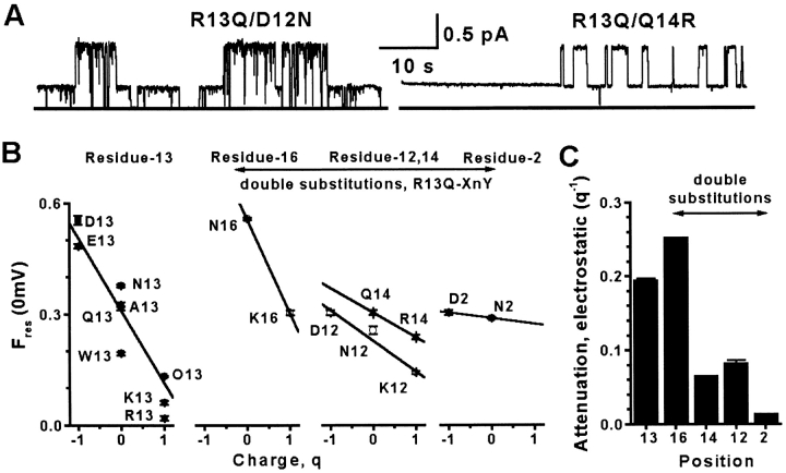
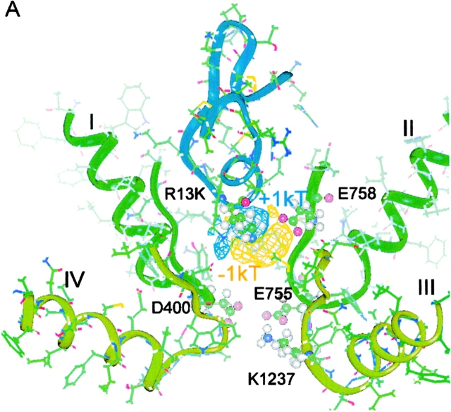
Pore-blocking toxins are valuable probes of ion channels that underlie electrical signaling. To be effective inhibitors, they must show high affinity and specificity and prevent ion conduction. The 22-residue sea snail peptide, mu-conotoxin GIIIA, blocks the skeletal muscle sodium channel completely. Partially blocking peptides, derived by making single or paired amino acid substitutions in mu-conotoxin GIIIA, allow a novel analysis of blocking mechanisms. Replacement of one critical residue (Arg-13) yielded peptides that only partially blocked single-channel current. These derivatives, and others with simultaneous substitution of a second residue, were used to elucidate the structural basis of the toxin's blocking action. The charge at residue-13 was the most striking determinant. A positive charge was necessary, though not sufficient, for complete block. Blocking efficacy increased with increasing residue-13 side chain size, regardless of charge, suggesting a steric contribution to inhibition. Charges grouped on one side of the toxin molecule at positions 2, 12, and 14 had a weaker influence, whereas residue-16, on the opposite face of the toxin, was more influential. Most directly interpreted, the data suggest that one side of the toxin is masked by close apposition to a binding surface on the pore, whereas the other side, bearing Lys-16, is exposed to an aqueous cavity accessible to entering ions. Strong charge-dependent effects emanate from this toxin surface. In the native toxin, Arg-13 probably presents a strategically placed electrostatic barrier rather than effecting a complete steric occlusion of the pore. This differs from other well-described channel inhibitors such as the charybdotoxin family of potassium channel blockers and the sodium channel-blocking guanidinium toxins (tetrodotoxin and saxitoxin), which appear to occlude the narrow part of the pore.
孔道阻断毒素是电信号传导所依赖的离子通道的重要探针。要成为有效的抑制剂,它们必须表现出高亲和力和特异性,并阻止离子传导。由22个氨基酸残基组成的海蜗牛肽——μ-芋螺毒素GIIIA,能完全阻断骨骼肌钠通道。通过对μ-芋螺毒素GIIIA进行单氨基酸或双氨基酸取代得到的部分阻断肽,可对阻断机制进行新的分析。替换一个关键残基(Arg-13)产生的肽只能部分阻断单通道电流。这些衍生物以及其他同时替换第二个残基的肽,被用于阐明毒素阻断作用的结构基础。残基13处的电荷是最显著的决定因素。正电荷对于完全阻断是必要的,但不是充分的。无论电荷如何,随着残基13侧链大小的增加,阻断效力增强,这表明空间因素对抑制作用有贡献。毒素分子一侧2、12和14位的电荷影响较弱,而毒素相对面上的残基16影响更大。最直接的解释是,数据表明毒素的一侧通过紧密贴合孔道上的结合表面而被掩盖,而另一侧带有Lys-16,暴露于可被进入离子接触的水腔中。这种毒素表面产生强烈的电荷依赖性效应。在天然毒素中,Arg-13可能呈现出一个策略性放置的静电屏障,而不是完全占据孔道的空间。这与其他已详细描述的通道抑制剂不同,如钾通道阻断剂的蝎毒素家族和钠通道阻断胍类毒素(河豚毒素和石房蛤毒素),它们似乎会阻塞孔道的狭窄部分。