Department of Physiology & Pharmacology, Cumming School of Medicine, University of Calgary, Calgary, AB T2N 4N1, Canada.
Illawarra Health and Medical Research Institute, University of Wollongong, Wollongong, NSW 2522, Australia.
Mar Drugs. 2019 Aug 29;17(9):510. doi: 10.3390/md17090510.
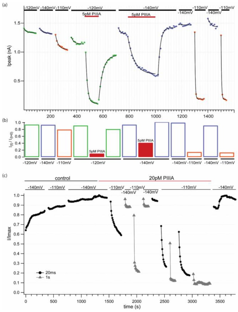
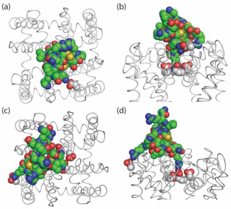
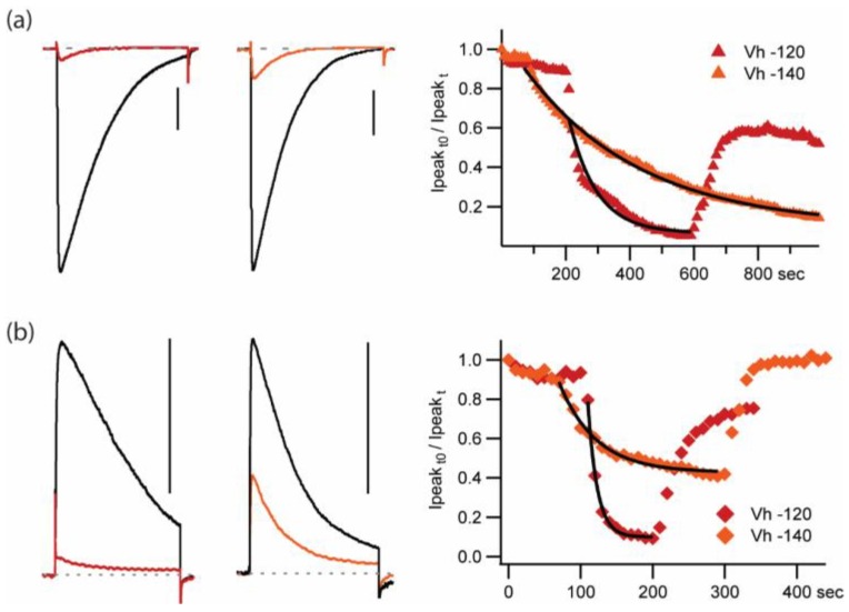
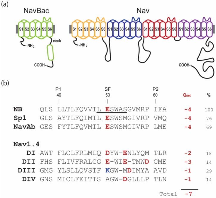
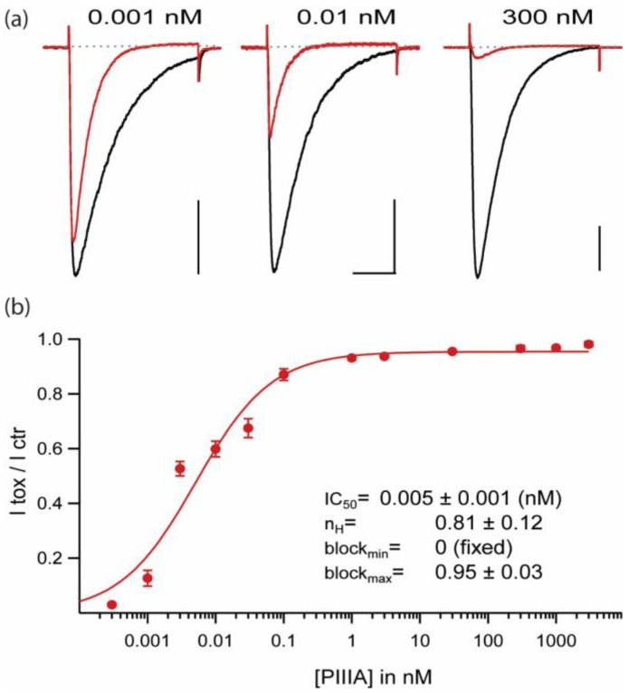
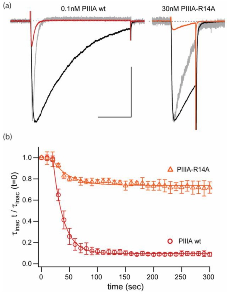
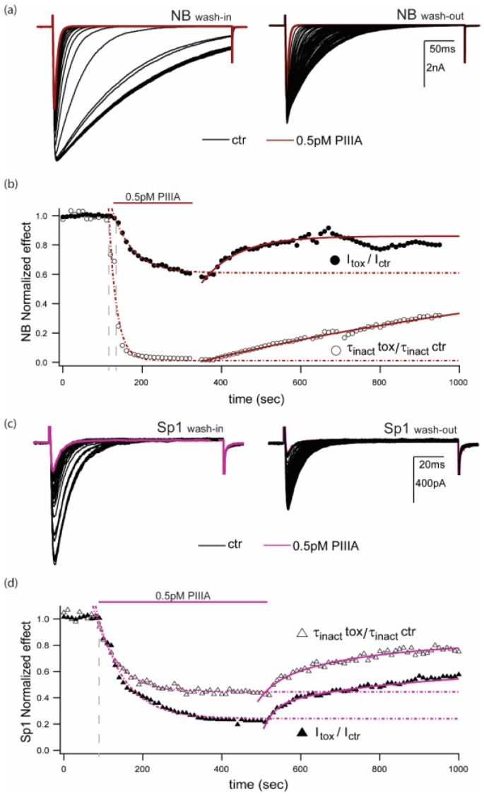
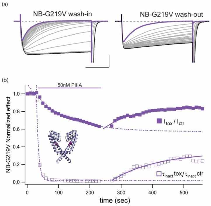
µ-Conotoxin PIIIA, in the sub-picomolar, range inhibits the archetypal bacterial sodium channel NaChBac (NavBh) in a voltage- and use-dependent manner. Peptide µ-conotoxins were first recognized as potent components of the venoms of fish-hunting cone snails that selectively inhibit voltage-gated skeletal muscle sodium channels, thus preventing muscle contraction. Intriguingly, computer simulations predicted that PIIIA binds to prokaryotic channel NavAb with much higher affinity than to fish (and other vertebrates) skeletal muscle sodium channel (Nav 1.4). Here, using whole-cell voltage clamp, we demonstrate that PIIIA inhibits NavBac mediated currents even more potently than predicted. From concentration-response data, with [PIIIA] varying more than 6 orders of magnitude (10 to 10 M), we estimated an IC = ~5 pM, maximal block of 0.95 and a Hill coefficient of 0.81 for the inhibition of peak currents. Inhibition was stronger at depolarized holding potentials and was modulated by the frequency and duration of the stimulation pulses. An important feature of the PIIIA action was acceleration of macroscopic inactivation. Docking of PIIIA in a NaChBac (NavBh) model revealed two interconvertible binding modes. In one mode, PIIIA sterically and electrostatically blocks the permeation pathway. In a second mode, apparent stabilization of the inactivated state was achieved by PIIIA binding between P2 helices and trans-membrane S5s from adjacent channel subunits, partially occluding the outer pore. Together, our experimental and computational results suggest that, besides blocking the channel-mediated currents by directly occluding the conducting pathway, PIIIA may also change the relative populations of conducting (activated) and non-conducting (inactivated) states.
µ-Conotoxin PIIIA 在皮摩尔范围内以电压和使用依赖性方式抑制典型的细菌钠通道 NaChBac(NavBh)。肽 µ-conotoxin 最初被认为是鱼猎圆锥蜗牛毒液中的有效成分,它们选择性地抑制电压门控骨骼肌钠离子通道,从而阻止肌肉收缩。有趣的是,计算机模拟预测 PIIIA 与原核通道 NavAb 的结合亲和力远高于鱼类(和其他脊椎动物)骨骼肌钠离子通道(Nav 1.4)。在这里,我们使用全细胞电压钳,证明 PIIIA 抑制 NavBac 介导的电流的能力比预测的更强。从浓度反应数据来看,[PIIIA]变化超过 6 个数量级(10 到 10 M),我们估计 IC = ~5 pM,对峰值电流的最大抑制为 0.95,Hill 系数为 0.81。在去极化保持电位下抑制作用更强,并受刺激脉冲频率和持续时间的调节。PIIIA 作用的一个重要特征是加速宏观失活。在 NaChBac(NavBh)模型中对接 PIIIA 显示了两种可相互转换的结合模式。在一种模式中,PIIIA 从立体和静电上阻断了渗透途径。在第二种模式中,PIIIA 结合在相邻通道亚基的 P2 螺旋和跨膜 S5 之间,实现了失活状态的明显稳定,部分阻塞了外孔。总之,我们的实验和计算结果表明,除了通过直接阻塞导电途径来阻断通道介导的电流外,PIIIA 还可能改变导电(激活)和非导电(失活)状态的相对比例。