McDonough Stefan I, Boland Linda M, Mintz Isabelle M, Bean Bruce P
Marine Biological Laboratory, Woods Hole, MA 02543, USA.
J Gen Physiol. 2002 Apr;119(4):313-28. doi: 10.1085/jgp.20028560.
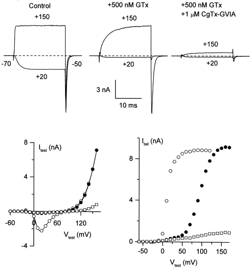
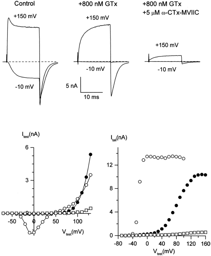
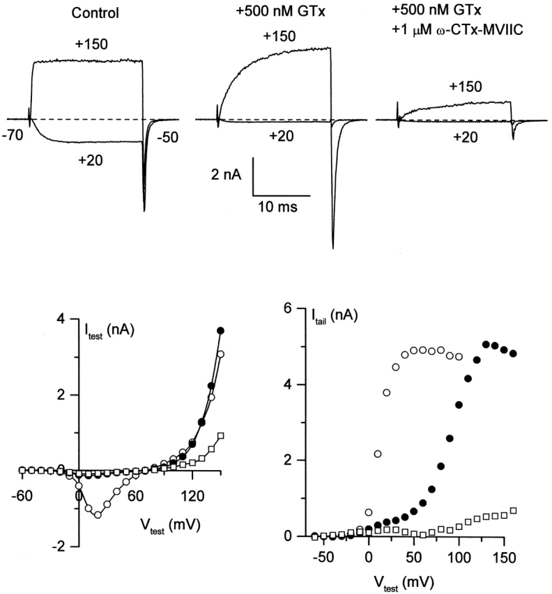
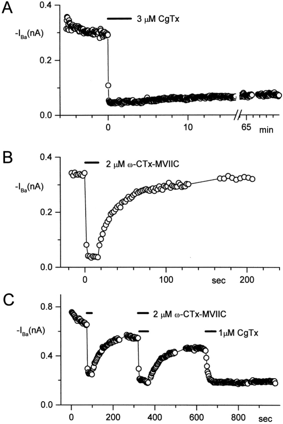
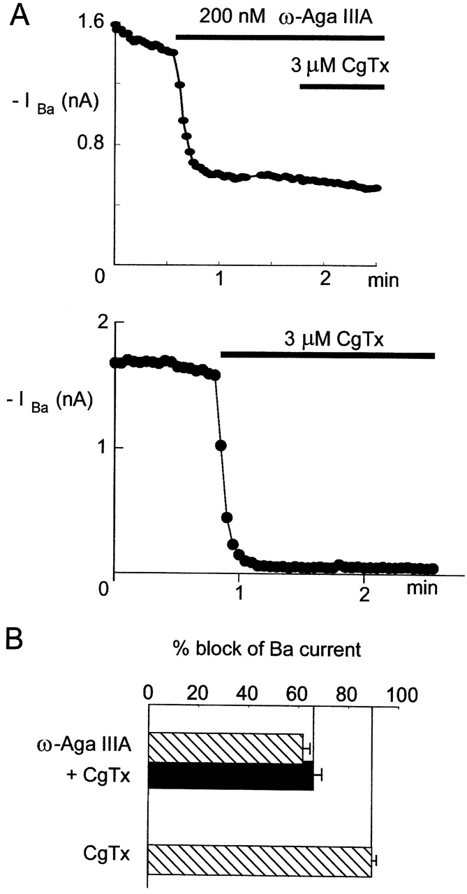
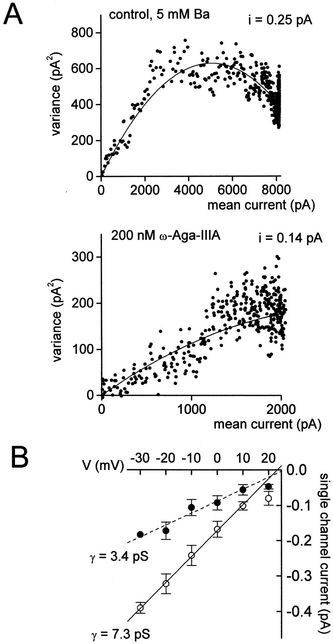
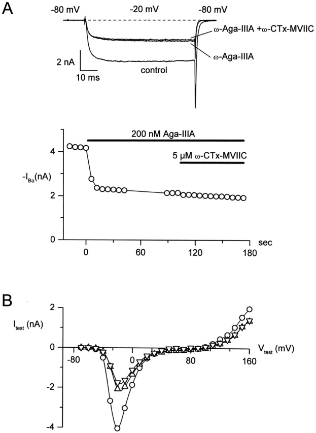
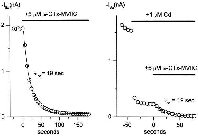
A number of peptide toxins from venoms of spiders and cone snails are high affinity ligands for voltage-gated calcium channels and are useful tools for studying calcium channel function and structure. Using whole-cell recordings from rat sympathetic ganglion and cerebellar Purkinje neurons, we studied toxins that target neuronal N-type (Ca(V)2.2) and P-type (Ca(V)2.1) calcium channels. We asked whether different toxins targeting the same channels bind to the same or different sites on the channel. Five toxins (omega-conotoxin-GVIA, omega-conotoxin MVIIC, omega-agatoxin-IIIA, omega-grammotoxin-SIA, and omega-agatoxin-IVA) were applied in pairwise combinations to either N- or P-type channels. Differences in the characteristics of inhibition, including voltage dependence, reversal kinetics, and fractional inhibition of current, were used to detect additive or mutually occlusive effects of toxins. Results suggest at least two distinct toxin binding sites on the N-type channel and three on the P-type channel. On N-type channels, results are consistent with blockade of the channel pore by omega-CgTx-GVIA, omega-Aga-IIIA, and omega-CTx-MVIIC, whereas grammotoxin likely binds to a separate region coupled to channel gating. omega-Aga-IIIA produces partial channel block by decreasing single-channel conductance. On P-type channels, omega-CTx-MVIIC and omega-Aga-IIIA both likely bind near the mouth of the pore. omega-Aga-IVA and grammotoxin each bind to distinct regions associated with channel gating that do not overlap with the binding region of pore blockers. For both N- and P-type channels, omega-CTx-MVIIC binding produces complete channel block, but is prevented by previous partial channel block by omega-Aga-IIIA, suggesting that omega-CTx-MVIIC binds closer to the external mouth of the pore than does omega-Aga-IIIA.
蜘蛛和芋螺毒液中的许多肽毒素是电压门控钙通道的高亲和力配体,是研究钙通道功能和结构的有用工具。利用大鼠交感神经节和小脑浦肯野神经元的全细胞记录,我们研究了靶向神经元N型(Ca(V)2.2)和P型(Ca(V)2.1)钙通道的毒素。我们询问靶向相同通道的不同毒素是否结合到通道上的相同或不同位点。将五种毒素(ω-芋螺毒素-GVIA、ω-芋螺毒素MVIIC、ω-阿加毒素-IIIA、ω-格拉莫毒素-SIA和ω-阿加毒素-IVA)以两两组合的方式应用于N型或P型通道。通过抑制特性的差异,包括电压依赖性、反转动力学和电流的分数抑制,来检测毒素的相加或相互闭塞效应。结果表明N型通道上至少有两个不同的毒素结合位点,P型通道上有三个。在N型通道上,结果与ω-CgTx-GVIA、ω-Aga-IIIA和ω-CTx-MVIIC对通道孔的阻断一致,而格拉莫毒素可能结合到与通道门控相关的单独区域。ω-Aga-IIIA通过降低单通道电导产生部分通道阻断。在P型通道上,ω-CTx-MVIIC和ω-Aga-IIIA都可能结合在孔口附近。ω-Aga-IVA和格拉莫毒素各自结合到与通道门控相关的不同区域,这些区域与孔道阻滞剂的结合区域不重叠。对于N型和P型通道,ω-CTx-MVIIC结合都会导致完全通道阻断,但之前由ω-Aga-IIIA产生的部分通道阻断会阻止这种情况,这表明ω-CTx-MVIIC比ω-Aga-IIIA更靠近孔的外口结合。