Gaillard Claire, Shlyakhtenko Luda S, Lyubchenko Yuri L, Strauss François
Institut Jacques Monod, 2 place Jussieu, 75251 Paris 05, France.
BMC Struct Biol. 2002 Nov 26;2:7. doi: 10.1186/1472-6807-2-7.
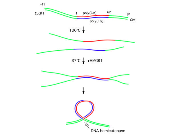
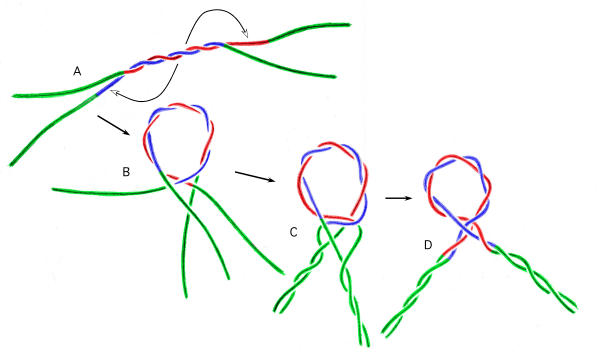
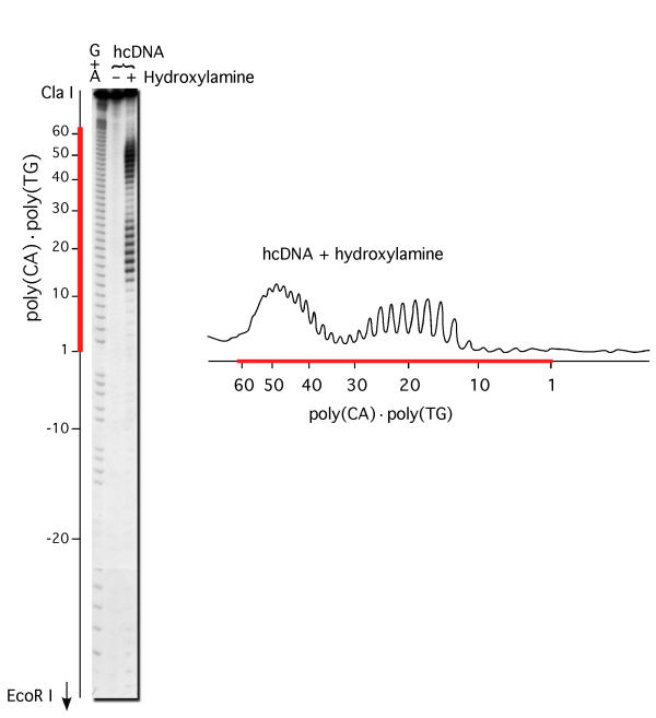
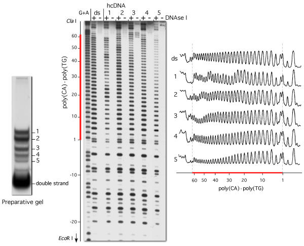
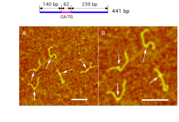
We have previously isolated a stable alternative DNA structure, which was formed in vitro by reassociation of the strands of DNA fragments containing a 62 bp tract of the CA-microsatellite poly(CA).poly(TG). In the model which was proposed for this structure the double helix is folded into a loop, the base of the loop consists of a DNA junction in which one of the strands of one duplex passes between the two strands of the other duplex, forming a DNA hemicatenane in a hemiknot structure. The hemiknot DNA structures obtained with long CA/TG inserts have been imaged by AFM allowing us to directly visualize the loops.
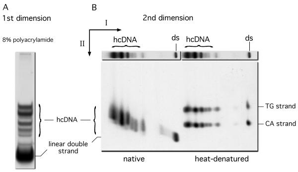
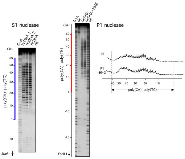
Here we have analyzed this structure with several different techniques: high-resolution gel electrophoresis, probing by digestion with single stranded DNA-specific nucleases or with DNase I, modification with chemicals specific for unpaired bases, and atomic force microscopy. The data show a change in DNA structure localized to the CA/TG sequence and allow us to better understand the structure of this alternative conformation and the mechanism of its formation.
The present work is in good agreement with the model of hemicatenated DNA loop proposed previously. In the presence of protein HMGB1, shifted reassociation of the strands of DNA fragments containing a tract of the poly(CA).poly(TG) microsatellite leads to the formation of DNA loops maintained at their base by a hemicatenated junction located within the repetitive sequence. No mobility of the junction along the DNA molecule could be detected under the conditions used. The novel possibility to prepare DNA hemicatenanes should be useful to further study this alternative DNA structure and its involvement in replication or recombination.
我们之前分离出了一种稳定的替代性DNA结构,它是在体外由包含62个碱基对的CA微卫星多聚(CA)·多聚(TG)的DNA片段链重新结合形成的。在为这种结构提出的模型中,双螺旋折叠成一个环,环的基部由一个DNA连接点组成,其中一个双链的一条链在另一个双链的两条链之间穿过,形成一个半结结构的DNA半连环体。用长CA/TG插入片段获得的半结DNA结构已通过原子力显微镜成像,使我们能够直接观察到环。
在这里,我们用几种不同的技术分析了这种结构:高分辨率凝胶电泳、用单链DNA特异性核酸酶或DNase I消化进行探测、用针对未配对碱基的化学物质进行修饰以及原子力显微镜。数据显示DNA结构的变化局限于CA/TG序列,使我们能够更好地理解这种替代构象的结构及其形成机制。
目前的工作与之前提出的半连环DNA环模型非常一致。在蛋白质HMGB1存在的情况下,含有多聚(CA)·多聚(TG)微卫星片段的DNA链的重新结合发生偏移,导致形成DNA环,其基部由位于重复序列内的半连环连接点维持。在所使用的条件下,未检测到连接点沿DNA分子的移动。制备DNA半连环体的新可能性对于进一步研究这种替代DNA结构及其在复制或重组中的作用应该是有用的。