• 文献检索
  • 文档翻译
  • 深度研究
  • 学术资讯
  • Suppr Zotero 插件Zotero 插件
  • 邀请有礼
  • 套餐&价格
  • 历史记录
应用&插件
Suppr Zotero 插件Zotero 插件浏览器插件Mac 客户端Windows 客户端微信小程序
定价
高级版会员购买积分包购买API积分包
服务
文献检索文档翻译深度研究API 文档MCP 服务
关于我们
关于 Suppr公司介绍联系我们用户协议隐私条款
关注我们

Suppr 超能文献

核心技术专利:CN118964589B侵权必究
粤ICP备2023148730 号-1Suppr @ 2026

文献检索

告别复杂PubMed语法,用中文像聊天一样搜索,搜遍4000万医学文献。AI智能推荐,让科研检索更轻松。

立即免费搜索

文件翻译

保留排版,准确专业,支持PDF/Word/PPT等文件格式,支持 12+语言互译。

免费翻译文档

深度研究

AI帮你快速写综述,25分钟生成高质量综述,智能提取关键信息,辅助科研写作。

立即免费体验

中间丝与膜的附着作用与肌动蛋白依赖性连接协同发挥功能,以调节细胞间黏附强度。

Intermediate filament-membrane attachments function synergistically with actin-dependent contacts to regulate intercellular adhesive strength.

作者信息

Huen Arthur C, Park Jung K, Godsel Lisa M, Chen Xuejun, Bannon Leslie J, Amargo Evangeline V, Hudson Tracie Y, Mongiu Anne K, Leigh Irene M, Kelsell David P, Gumbiner Barry M, Green Kathleen J

机构信息

Department of Pathology, Northwestern University Feinberg School of Medicine, Chicago, IL 60611, USA.

出版信息

J Cell Biol. 2002 Dec 23;159(6):1005-17. doi: 10.1083/jcb.200206098.

DOI:10.1083/jcb.200206098
PMID:12499357
原文链接:https://pmc.ncbi.nlm.nih.gov/articles/PMC2173978/
Abstract

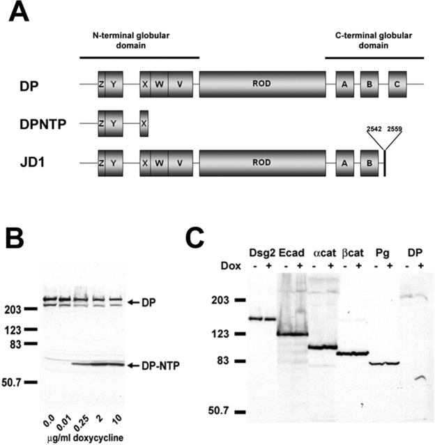
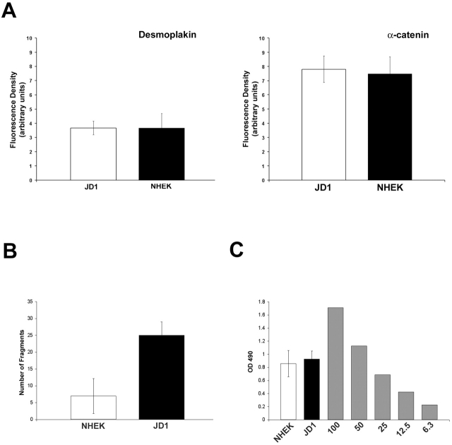
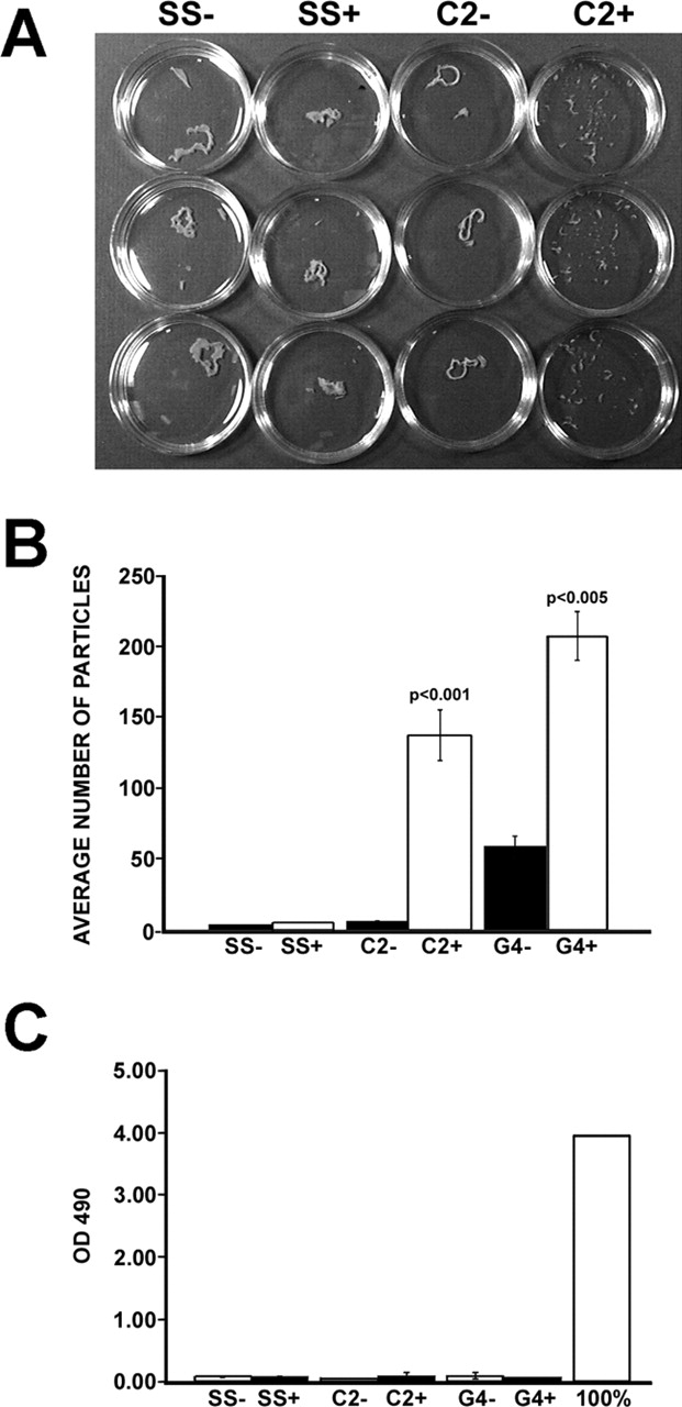
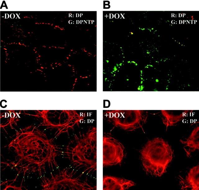
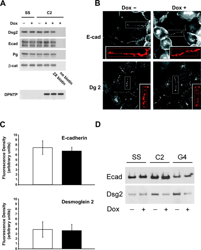
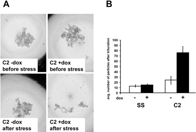
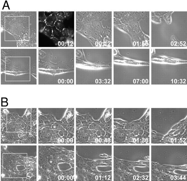
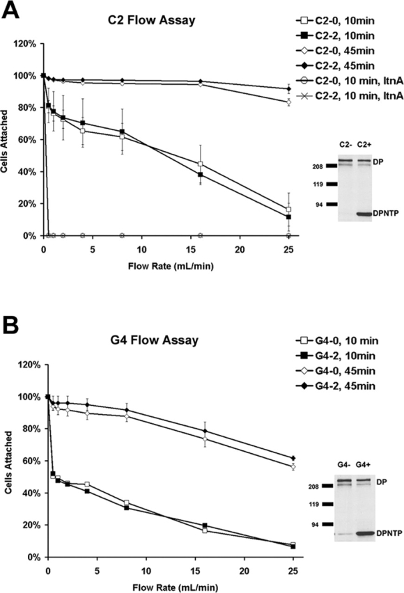
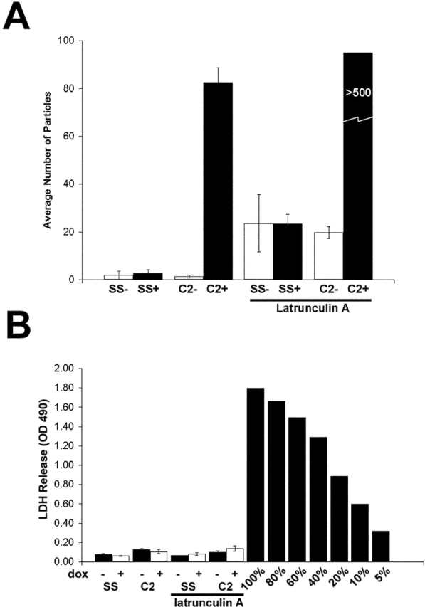
By tethering intermediate filaments (IFs) to sites of intercellular adhesion, desmosomes facilitate formation of a supercellular scaffold that imparts mechanical strength to a tissue. However, the role IF-membrane attachments play in strengthening adhesion has not been directly examined. To address this question, we generated Tet-On A431 cells inducibly expressing a desmoplakin (DP) mutant lacking the rod and IF-binding domains (DPNTP). DPNTP localized to the plasma membrane and led to dissociation of IFs from the junctional plaque, without altering total or cell surface distribution of adherens junction or desmosomal proteins. However, a specific decrease in the detergent-insoluble pool of desmoglein suggested a reduced association with the IF cytoskeleton. DPNTP-expressing cell aggregates in suspension or substrate-released cell sheets readily dissociated when subjected to mechanical stress whereas controls remained largely intact. Dissociation occurred without lactate dehydrogenase release, suggesting that loss of tissue integrity was due to reduced adhesion rather than increased cytolysis. JD-1 cells from a patient with a DP COOH-terminal truncation were also more weakly adherent compared with normal keratinocytes. When used in combination with DPNTP, latrunculin A, which disassembles actin filaments and disrupts adherens junctions, led to dissociation up to an order of magnitude greater than either treatment alone. These data provide direct in vitro evidence that IF-membrane attachments regulate adhesive strength and suggest furthermore that actin- and IF-based junctions act synergistically to strengthen adhesion.

摘要

通过将中间丝(IFs)拴系到细胞间粘附位点,桥粒促进了超细胞支架的形成,该支架赋予组织机械强度。然而,IF-膜附着在增强粘附中所起的作用尚未得到直接研究。为了解决这个问题,我们构建了可诱导表达缺乏杆状和IF结合结构域的桥粒斑蛋白(DP)突变体(DPNTP)的Tet-On A431细胞。DPNTP定位于质膜,导致IFs从连接斑解离,而不改变黏附连接或桥粒蛋白的总量或细胞表面分布。然而,桥粒芯糖蛋白的去污剂不溶性组分的特异性降低表明与IF细胞骨架的结合减少。表达DPNTP的细胞聚集体在悬浮液中或从底物释放的细胞片在受到机械应力时容易解离,而对照基本保持完整。解离发生时没有乳酸脱氢酶释放,表明组织完整性的丧失是由于粘附力降低而不是细胞溶解增加。来自一名患有DP羧基末端截短的患者的JD-1细胞与正常角质形成细胞相比,粘附力也更弱。当与DPNTP联合使用时,破坏肌动蛋白丝并破坏黏附连接的拉春库林A导致的解离比单独任何一种处理大一个数量级。这些数据提供了直接的体外证据,表明IF-膜附着调节粘附强度,并且进一步表明基于肌动蛋白和IF的连接协同作用以增强粘附。

https://cdn.ncbi.nlm.nih.gov/pmc/blobs/3cba/2173978/e64e3fd6330e/200206098f9.jpg
https://cdn.ncbi.nlm.nih.gov/pmc/blobs/3cba/2173978/a0435fdc7598/200206098f1.jpg
https://cdn.ncbi.nlm.nih.gov/pmc/blobs/3cba/2173978/efcf406406e6/200206098f5.jpg
https://cdn.ncbi.nlm.nih.gov/pmc/blobs/3cba/2173978/6c1efc119de7/200206098f2.jpg
https://cdn.ncbi.nlm.nih.gov/pmc/blobs/3cba/2173978/5086634b926a/200206098f3.jpg
https://cdn.ncbi.nlm.nih.gov/pmc/blobs/3cba/2173978/7fb07305db5b/200206098f4.jpg
https://cdn.ncbi.nlm.nih.gov/pmc/blobs/3cba/2173978/6b20f0c67648/200206098f6ab.jpg
https://cdn.ncbi.nlm.nih.gov/pmc/blobs/3cba/2173978/8a7d4c591aba/200206098f7.jpg
https://cdn.ncbi.nlm.nih.gov/pmc/blobs/3cba/2173978/a9fd0f905ca3/200206098f8.jpg
https://cdn.ncbi.nlm.nih.gov/pmc/blobs/3cba/2173978/e64e3fd6330e/200206098f9.jpg

相似文献

1
Intermediate filament-membrane attachments function synergistically with actin-dependent contacts to regulate intercellular adhesive strength.中间丝与膜的附着作用与肌动蛋白依赖性连接协同发挥功能,以调节细胞间黏附强度。
J Cell Biol. 2002 Dec 23;159(6):1005-17. doi: 10.1083/jcb.200206098.
2
Regulated expression of human filaggrin in keratinocytes results in cytoskeletal disruption, loss of cell-cell adhesion, and cell cycle arrest.人丝聚合蛋白在角质形成细胞中的调控表达导致细胞骨架破坏、细胞间黏附丧失和细胞周期停滞。
Exp Cell Res. 2001 Nov 1;270(2):199-213. doi: 10.1006/excr.2001.5348.
3
Breaking the connection: displacement of the desmosomal plaque protein desmoplakin from cell-cell interfaces disrupts anchorage of intermediate filament bundles and alters intercellular junction assembly.切断连接:桥粒斑蛋白桥粒芯蛋白从细胞-细胞界面的位移破坏了中间丝束的锚定并改变了细胞间连接组装。
J Cell Biol. 1996 Aug;134(4):985-1001. doi: 10.1083/jcb.134.4.985.
4
Desmosomes and Intermediate Filaments: Their Consequences for Tissue Mechanics.桥粒与中间丝:它们对组织力学的影响
Cold Spring Harb Perspect Biol. 2017 Jun 1;9(6):a029157. doi: 10.1101/cshperspect.a029157.
5
Identification of the plakoglobin-binding domain in desmoglein and its role in plaque assembly and intermediate filament anchorage.桥粒芯糖蛋白中桥粒斑珠蛋白结合结构域的鉴定及其在斑块组装和中间丝锚定中的作用。
J Cell Biol. 1994 Oct;127(1):151-60. doi: 10.1083/jcb.127.1.151.
6
Striate palmoplantar keratoderma arising from desmoplakin and desmoglein 1 mutations is associated with contrasting perturbations of desmosomes and the keratin filament network.由桥粒斑蛋白和桥粒芯糖蛋白1突变引起的线状掌跖角化病与桥粒和角蛋白丝网络的对比性扰动有关。
Br J Dermatol. 2004 May;150(5):878-91. doi: 10.1111/j.1365-2133.2004.05996.x.
7
Structure and function of desmosomal transmembrane core and plaque molecules.桥粒跨膜核心和斑块分子的结构与功能
Biophys Chem. 1994 May;50(1-2):97-112. doi: 10.1016/0301-4622(94)85023-2.
8
Different effects of dominant negative mutants of desmocollin and desmoglein on the cell-cell adhesion of keratinocytes.桥粒芯蛋白和桥粒芯胶粘蛋白的显性负性突变体对角质形成细胞细胞间黏附的不同影响。
J Cell Sci. 2000 May;113 ( Pt 10):1803-11. doi: 10.1242/jcs.113.10.1803.
9
Plakophilin 1 interferes with plakoglobin binding to desmoplakin, yet together with plakoglobin promotes clustering of desmosomal plaque complexes at cell-cell borders.桥粒芯蛋白1干扰桥粒斑珠蛋白与桥粒斑蛋白的结合,但与桥粒斑珠蛋白一起促进桥粒斑块复合物在细胞-细胞边界处聚集。
J Cell Sci. 2001 Feb;114(Pt 4):727-38. doi: 10.1242/jcs.114.4.727.
10
Plakoglobin is required for effective intermediate filament anchorage to desmosomes.桥粒斑珠蛋白是中间丝有效锚定于桥粒所必需的。
J Invest Dermatol. 2008 Nov;128(11):2665-2675. doi: 10.1038/jid.2008.141. Epub 2008 May 22.

引用本文的文献

1
Data-driven image analysis to determine antibody-induced dissociation of cell-cell adhesion and antibody pathogenicity in pemphigus.数据驱动的图像分析,用于确定天疱疮中抗体诱导的细胞间粘附解离和抗体致病性。
PNAS Nexus. 2025 Jul 18;4(8):pgaf218. doi: 10.1093/pnasnexus/pgaf218. eCollection 2025 Aug.
2
Pemphigus Vulgaris Autoantibodies Induce an Endoplasmic Reticulum Stress Response.寻常型天疱疮自身抗体诱导内质网应激反应。
J Invest Dermatol. 2025 Feb 3. doi: 10.1016/j.jid.2024.12.028.
3
The transmembrane domain of the desmosomal cadherin desmoglein-1 governs lipid raft association to promote desmosome adhesive strength.

本文引用的文献

1
F-actin serves as a template for cytokeratin organization in cell free extracts.F-肌动蛋白在无细胞提取物中作为细胞角蛋白组织的模板。
J Cell Sci. 2002 Apr 1;115(Pt 7):1373-82. doi: 10.1242/jcs.115.7.1373.
2
Cadherin-directed actin assembly: E-cadherin physically associates with the Arp2/3 complex to direct actin assembly in nascent adhesive contacts.钙黏蛋白介导的肌动蛋白组装:E-钙黏蛋白与Arp2/3复合体发生物理结合,以在新生黏附接触点引导肌动蛋白组装。
Curr Biol. 2002 Mar 5;12(5):379-82. doi: 10.1016/s0960-9822(02)00661-9.
3
'Hard' and 'soft' principles defining the structure, function and regulation of keratin intermediate filaments.
桥粒黏附分子 desmoglein-1 的跨膜结构域调控脂筏结合以增强桥粒黏附力。
Mol Biol Cell. 2024 Dec 1;35(12):ar152. doi: 10.1091/mbc.E24-05-0200. Epub 2024 Nov 6.
4
Data-Driven Image Analysis to Determine Antibody-Induced Dissociation of Cell-Cell Adhesion and Antibody Pathogenicity in Pemphigus Vulgaris.基于数据驱动的图像分析以确定寻常型天疱疮中抗体诱导的细胞间黏附解离及抗体致病性
bioRxiv. 2024 Oct 13:2024.10.09.617446. doi: 10.1101/2024.10.09.617446.
5
The desmosome-intermediate filament system facilitates mechanotransduction at adherens junctions for epithelial homeostasis.桥粒-中间丝系统有助于细胞连接黏着斑处的机械转导,从而维持上皮组织稳态。
Curr Biol. 2024 Sep 9;34(17):4081-4090.e5. doi: 10.1016/j.cub.2024.07.074. Epub 2024 Aug 16.
6
Targeting SERCA2 in organotypic epidermis reveals MEK inhibition as a therapeutic strategy for Darier disease.靶向器官型表皮中的 SERCA2 揭示了 MEK 抑制作为 Darier 病的一种治疗策略。
JCI Insight. 2023 Sep 22;8(18):e170739. doi: 10.1172/jci.insight.170739.
7
Towards a Better Understanding of Genotype-Phenotype Correlations and Therapeutic Targets for Cardiocutaneous Genes: The Importance of Functional Studies above Prediction.为了更好地理解基因型-表型相关性和心脏皮肤基因的治疗靶点:功能研究优于预测的重要性。
Int J Mol Sci. 2022 Sep 15;23(18):10765. doi: 10.3390/ijms231810765.
8
The β-Subunit (AMOG) of Human Na, K-ATPase Is a Homophilic Adhesion Molecule.人源 Na, K-ATPase 的β亚基(AMOG)是一种同亲性黏附分子。
Int J Mol Sci. 2022 Jul 14;23(14):7753. doi: 10.3390/ijms23147753.
9
Pitfalls in the Application of Dispase-Based Keratinocyte Dissociation Assay for In Vitro Analysis of Pemphigus Vulgaris.基于Dispase的角质形成细胞解离试验在寻常型天疱疮体外分析中的应用陷阱
Vaccines (Basel). 2022 Jan 28;10(2):208. doi: 10.3390/vaccines10020208.
10
Differential Pathomechanisms of Desmoglein 1 Transmembrane Domain Mutations in Skin Disease.皮肤疾病中桥粒芯糖蛋白 1 跨膜结构域突变的差异发病机制。
J Invest Dermatol. 2022 Feb;142(2):323-332.e8. doi: 10.1016/j.jid.2021.07.154. Epub 2021 Aug 2.
定义角蛋白中间丝结构、功能和调节的“硬”和“软”原则。
Curr Opin Cell Biol. 2002 Feb;14(1):110-22. doi: 10.1016/s0955-0674(01)00301-5.
4
Deconstructing desmoplakin.解构桥粒斑蛋白
Nat Cell Biol. 2001 Dec;3(12):E270-2. doi: 10.1038/ncb1201-e270.
5
Desmoplakin is essential in epidermal sheet formation.桥粒斑蛋白在表皮片层形成中至关重要。
Nat Cell Biol. 2001 Dec;3(12):1076-85. doi: 10.1038/ncb1201-1076.
6
The plakin family.斑联蛋白家族。
J Cell Sci. 2001 Oct;114(Pt 19):3409-10. doi: 10.1242/jcs.114.19.3409.
7
Desmosomes: structure and function in normal and diseased epidermis.桥粒:正常及病变表皮中的结构与功能
J Dermatol. 2001 Jun;28(6):291-8. doi: 10.1111/j.1346-8138.2001.tb00136.x.
8
E-cadherin suppresses cellular transformation by inhibiting beta-catenin signaling in an adhesion-independent manner.E-钙黏蛋白通过以一种不依赖黏附的方式抑制β-连环蛋白信号传导来抑制细胞转化。
J Cell Biol. 2001 May 28;153(5):1049-60. doi: 10.1083/jcb.153.5.1049.
9
Tyrosine-phosphorylated plakoglobin is associated with desmogleins but not desmoplakin after epidermal growth factor receptor activation.表皮生长因子受体激活后,酪氨酸磷酸化的桥粒芯蛋白与桥粒芯糖蛋白相关,但与桥粒斑蛋白无关。
J Biol Chem. 2001 Jul 6;276(27):24871-80. doi: 10.1074/jbc.M102731200. Epub 2001 May 2.
10
Are desmosomes more than tethers for intermediate filaments?桥粒仅仅是中间丝的连接结构吗?
Nat Rev Mol Cell Biol. 2000 Dec;1(3):208-16. doi: 10.1038/35043032.