Araujo-Nascimento M d, Désormeaux Y, Cantin M
Am J Pathol. 1976 Mar;82(3):527-48.
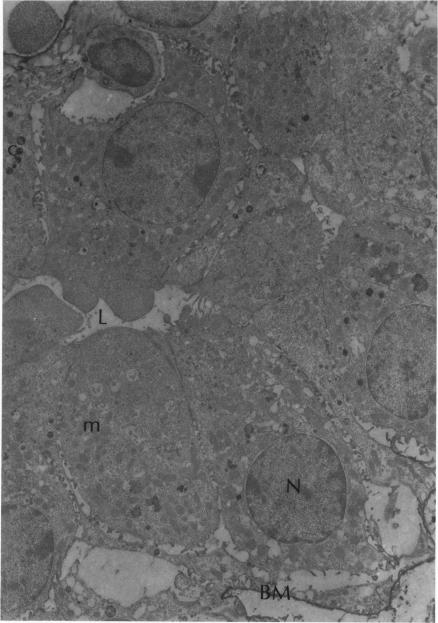
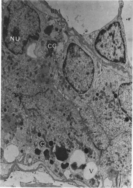
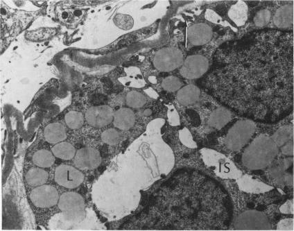
Partial ligation of the aorta between the renal arteries induces marked atrophy of the cortical tubules (including the macula densa) of the left (endocrine) kidney with a remarkable increase in the number and granularity of hypersecretory juxtaglomerular granulated cells (JGC) which are found not only at the glomerular pole of arterioles but also in the walls of arteries and arterioles far removed from the glomerulus. Staining of fine sections of Araldite-embedded endocrine kidneys according to the periodic acid-thiocarbohydrazide-silver proteinate technique of Thiery reveals abundant glycogen in the JGC and less in the blood vessels and tubules. Juxtaglomerular granules are argentaphobic, but their rim is positively stained when ultrathin sections of glutaraldehyde-fixed, glycol methacrylate-embedded kidneys are exposed to phosphotungstic acid at a low pH. A positive reaction is also shown by the cell coat and lysosomes of JGC as well as by the thickened basal lamina, cell coat, cytosomes, and cytosegresomes of the atrophic tubules. Atrophy is most pronounced in the proximal convoluted tubules, which lose their apical microvilli, their basal infoldings and the majority of their mitochondria and cytosomes.
在肾动脉之间对主动脉进行部分结扎,会导致左(内分泌)肾的皮质小管(包括致密斑)显著萎缩,同时,高分泌性肾小球旁颗粒细胞(JGC)的数量和颗粒度显著增加,这些细胞不仅存在于小动脉的肾小球极,还存在于远离肾小球的动脉和小动脉壁中。根据蒂埃里的高碘酸 - 硫代碳酰肼 - 银蛋白技术,对用环氧树脂包埋的内分泌肾的薄切片进行染色,结果显示JGC中糖原丰富,而血管和小管中的糖原较少。肾小球旁颗粒对嗜银染色呈阴性,但当用戊二醛固定、甲基丙烯酸乙二醇酯包埋的肾脏超薄切片在低pH值下暴露于磷钨酸时,其边缘呈阳性染色。JGC的细胞被膜和溶酶体以及萎缩小管的增厚基膜、细胞被膜、胞质体和胞质分泌体也显示出阳性反应。萎缩在近端曲管中最为明显,近端曲管会失去其顶端微绒毛、基底褶皱以及大部分线粒体和胞质体。