Cantin M, Araujo-Nascimento M D, Benchimol S, Desormeaux Y
Am J Pathol. 1977 Jun;87(3):581-602.
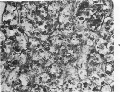
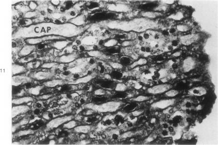
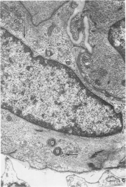
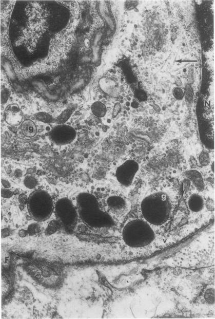
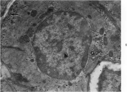
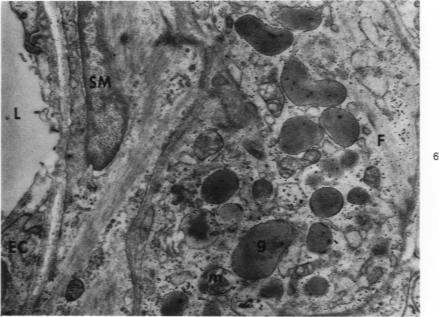
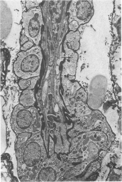
Partial ligation of the aorta between the renal arteries induces marked atrophy of the cortical tubules of the left (endocrine) kidney with a remarkable increase in the number and granularity of hypersecretory juxtaglomerular cells (JGC), which are found not only at the glomerular pole of arterioles but also in the walls of arteries and arterioles far removed from the glomerulus. Typical vascular smooth muscle cells (SMC), in which secretory granules appear, show a concomitant development of their Golgi complex and rough endoplasmic reticulum, with a gradual decrease in the number of their filaments. Microtubules also appear in the Golgi area. Thiery's periodic acid-thiocarbohydrazide-silver proteinate technique demonstrates that in these "intermediate" cells, as in mature JGC, the amount of glycogen is greater than in SMC. The newly-developed secretory granules of intermediate cells are stained by phosphotungstic acid at a low pH, as are the mature granules of JGC, an indication that both types contain glycoproteins. Light and electron microscopic autoradiography reveal that both JGC and "intermediate" cells of the vascular wall do not incorporate radioactive thymidine (injected during the 10-day observation period). Thus, they develop by metaplasia of preexistent SMC. In control kidneys, radioactive thymidine is practically never incorporated into the nuclei of SMC but is found in a few glomerular and tubular cells of all zones except the papilla.The endocrine kidney shows virtually no reactive nuclei in vascular SMC, glomeruli, or tubular cells of the outer cortex. Thymidine is incorporated into practically all nuclei of the straight portion of proximal tubules and into about half the nuclei of all medullary tubular cells including the papilla.
在肾动脉之间对主动脉进行部分结扎,会导致左(内分泌)肾皮质小管显著萎缩,同时分泌亢进的球旁细胞(JGC)数量和颗粒度显著增加,这些细胞不仅存在于小动脉的肾小球极,还存在于远离肾小球的动脉和小动脉壁中。典型的血管平滑肌细胞(SMC)出现分泌颗粒,其高尔基体复合体和粗面内质网随之发育,细丝数量逐渐减少。微管也出现在高尔基体区域。蒂埃里的高碘酸 - 硫代碳酰肼 - 银蛋白技术表明,在这些“中间”细胞中,如同在成熟的JGC中一样,糖原含量高于SMC。中间细胞新形成的分泌颗粒在低pH值下被磷钨酸染色,JGC的成熟颗粒也是如此,这表明两种类型的颗粒都含有糖蛋白。光镜和电镜放射自显影显示,血管壁的JGC和“中间”细胞均不摄取放射性胸苷(在10天观察期内注射)。因此,它们是由预先存在的SMC化生而来。在对照肾脏中,放射性胸苷实际上从未进入SMC的细胞核,但在除乳头外所有区域的少数肾小球和肾小管细胞中可以发现。内分泌肾在外皮质的血管SMC、肾小球或肾小管细胞中几乎没有反应性细胞核。胸苷几乎被纳入近端小管直部的所有细胞核以及包括乳头在内的所有髓质肾小管细胞约一半的细胞核中。