Lénárt Péter, Rabut Gwénaël, Daigle Nathalie, Hand Arthur R, Terasaki Mark, Ellenberg Jan
Gene Expression and Cell Biology/Biophysics Programmes, European Molecular Biology Laboratory, D-69117 Heidelberg, Germany.
J Cell Biol. 2003 Mar 31;160(7):1055-68. doi: 10.1083/jcb.200211076. Epub 2003 Mar 24.
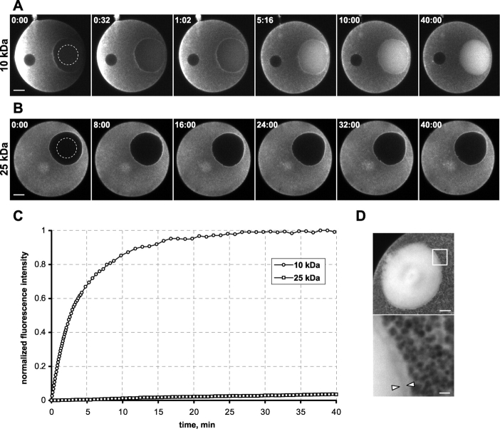
Breakdown of the nuclear envelope (NE) was analyzed in live starfish oocytes using a size series of fluorescently labeled dextrans, membrane dyes, and GFP-tagged proteins of the nuclear pore complex (NPC) and the nuclear lamina. Permeabilization of the nucleus occurred in two sequential phases. In phase I the NE became increasingly permeable for molecules up to approximately 40 nm in diameter, concurrent with a loss of peripheral nuclear pore components over a time course of 10 min. The NE remained intact on the ultrastructural level during this time. In phase II the NE was completely permeabilized within 35 s. This rapid permeabilization spread as a wave from one epicenter on the animal half across the nuclear surface and allowed free diffusion of particles up to approximately 100 nm in diameter into the nucleus. While the lamina and nuclear membranes appeared intact at the light microscopic level, a fenestration of the NE was clearly visible by electron microscopy in phase II. We conclude that NE breakdown in starfish oocytes is triggered by slow sequential disassembly of the NPCs followed by a rapidly spreading fenestration of the NE caused by the removal of nuclear pores from nuclear membranes still attached to the lamina.
利用一系列不同大小的荧光标记葡聚糖、膜染料以及核孔复合体(NPC)和核纤层的绿色荧光蛋白标记蛋白,对活海星卵母细胞中的核膜(NE)解体进行了分析。细胞核的通透化过程分为两个连续阶段。在第一阶段,核膜对直径约40纳米以下的分子通透性逐渐增加,同时在10分钟的时间进程中,外周核孔成分逐渐丢失。在此期间,核膜在超微结构水平上保持完整。在第二阶段,核膜在35秒内完全通透化。这种快速通透化以波的形式从动物极的一个震中穿过核表面传播,使得直径约100纳米以下的颗粒能够自由扩散到细胞核中。虽然在光学显微镜下核纤层和核膜看起来是完整的,但在第二阶段通过电子显微镜可以清楚地看到核膜有窗孔。我们得出结论,海星卵母细胞中的核膜解体是由NPC的缓慢顺序拆卸引发的,随后是由于从仍附着在核纤层上的核膜上去除核孔而导致的核膜快速扩展窗孔。