Yamamoto Y, Klein T W, Brown K, Friedman H
Department of Medical Microbiology and Immunology, University of South Florida College of Medicine, Tampa 33612.
Infect Immun. 1992 Aug;60(8):3231-7. doi: 10.1128/iai.60.8.3231-3237.1992.
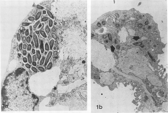
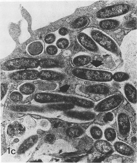
Legionella pneumophila infection of macrophages from permissive guinea pigs and from A/J mice compared with infection of cells from nonpermissive BDF1 mice was studied by electron microscopy. The cells from the BDF1 mice were nonpermissive for legionella growth in vitro and showed few if any bacteria in phagosomes by electron microscopic examination. Similar electron micrographic examination of macrophages from A/J mice permissive for legionella growth showed numerous intact intracellular bacteria within 24 to 48 h of culture and the transition of intracellular bacteria from localization in a few large vacuoles early in the course of infection to later localization in areas surrounded and studded by ribosomes. These electron microscopic observations were similar to those seen in the case of guinea pig macrophages infected with legionellae. Biochemical studies of macrophages from permissive versus nonpermissive animals showed little or no differences in respiratory burst and lysosomal enzyme activity for macrophages from all animals tested. However, when zymosan was used as a stimulant, macrophages from the nonpermissive mouse strain produced a larger amount of H2O2 and O2- than did cells from permissive guinea pigs or A/J mice. However, legionella vaccine itself induced no detectable or very little H2O2 and O2- in macrophages tested from any source. These results suggest that permissiveness of A/J mouse macrophages to legionella growth may involve mechanisms similar to those occurring in guinea pig macrophages in terms of morphologic and possibly even biochemical events. The relatively higher production of reactive oxygens by BDF1 mouse macrophages in response to zymosan correlated with nonpermissiveness for legionella growth, although further analysis is necessary to link these observations.
通过电子显微镜研究了嗜肺军团菌对来自易感性豚鼠和A/J小鼠的巨噬细胞的感染,并与对来自非易感性BDF1小鼠的细胞的感染进行了比较。BDF1小鼠的细胞在体外对军团菌生长不敏感,通过电子显微镜检查发现吞噬体中几乎没有细菌。对允许军团菌生长的A/J小鼠的巨噬细胞进行类似的电子显微镜检查,结果显示在培养24至48小时内有大量完整的细胞内细菌,并且细胞内细菌从感染初期在少数大液泡中的定位转变为后期在被核糖体包围和点缀的区域中的定位。这些电子显微镜观察结果与感染军团菌的豚鼠巨噬细胞的观察结果相似。对来自易感性与非易感性动物的巨噬细胞的生化研究表明,所有测试动物的巨噬细胞在呼吸爆发和溶酶体酶活性方面几乎没有差异。然而,当使用酵母聚糖作为刺激物时,来自非易感性小鼠品系的巨噬细胞比来自易感性豚鼠或A/J小鼠的细胞产生更多的H2O2和O2-。然而,军团菌疫苗本身在任何来源的测试巨噬细胞中均未诱导可检测到的或非常少量的H2O2和O2-。这些结果表明,A/J小鼠巨噬细胞对军团菌生长的易感性可能在形态学甚至可能在生化事件方面涉及与豚鼠巨噬细胞中发生的机制相似的机制。BDF1小鼠巨噬细胞对酵母聚糖反应产生的活性氧相对较高,这与对军团菌生长的非易感性相关,尽管需要进一步分析来将这些观察结果联系起来。