Nikol S, Isner J M, Pickering J G, Kearney M, Leclerc G, Weir L
Department of Medicine (Cardiology), St. Elizabeth's Hospital, Tufts University School of Medicine, Boston, Massachusetts 02135.
J Clin Invest. 1992 Oct;90(4):1582-92. doi: 10.1172/JCI116027.
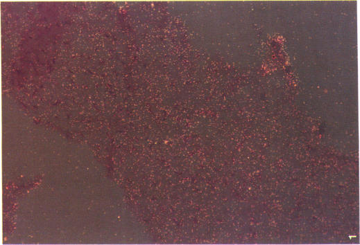
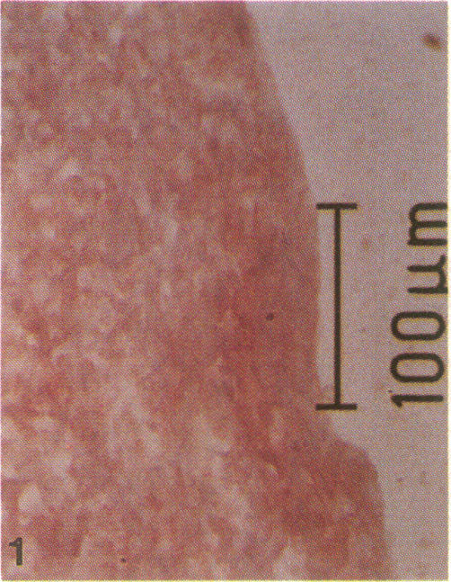
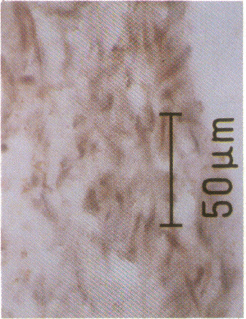
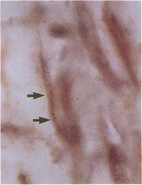
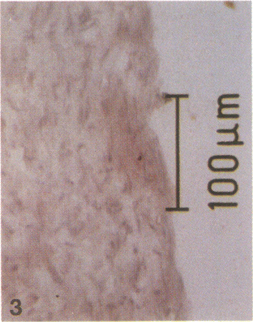
Human atheromata obtained in vivo were used to test the hypothesis that transforming growth factor-beta 1 plays a role in the development of vascular restenosis. We analyzed 28 specimens from patients with primary atherosclerotic or restenotic lesions; 26 of these were obtained by directional atherectomy and 2 at the time of coronary bypass surgery. Seven control tissues included operatively excised segments of human internal mammary artery, myocardium, and unused portions of vein graft obtained intraoperatively. From these 35 specimens, 210 sections were examined using in situ hybridization. Measurement of silver grains/nucleus disclosed that expression of transforming growth factor-beta 1 mRNA was highest in restenotic tissues (P < 0.001 vs. primary atherosclerotic tissues) and lowest in nonatherosclerotic (control) tissues. In cultures of human vascular smooth muscle cells grown from explants of internal mammary artery, expression of mRNA for transforming growth factor-beta 1 was significantly greater in subconfluent than in confluent smooth muscle cells (P = 0.05). Transforming growth factor type-beta III receptor was expressed in cell cultures and undetectable in the tissue specimens. Sections taken adjacent to those studied by in situ hybridization were examined by immunohistochemistry using antibodies against transforming growth factor-beta 1 and alpha-actin (as a marker for smooth muscle cells) and disclosed transforming growth factor-beta 1 in smooth muscle cells present in these sections. These findings are consistent with the concept that transforming growth factor-beta 1 plays an important role in modulating repair of vascular injury, including restenosis, after balloon angioplasty.
采用取自体内的人类动脉粥样硬化斑块来检验转化生长因子β1在血管再狭窄发展过程中起作用这一假说。我们分析了28例原发性动脉粥样硬化或再狭窄病变患者的标本;其中26例通过定向斑块旋切术获取,2例在冠状动脉搭桥手术时获取。7个对照组织包括术中切除的人类乳内动脉节段、心肌以及术中获取的未使用的静脉移植物部分。从这35个标本中,制备了210个切片用于原位杂交检测。银颗粒/细胞核计数显示,转化生长因子β1 mRNA在再狭窄组织中的表达最高(与原发性动脉粥样硬化组织相比,P < 0.001),在非动脉粥样硬化(对照)组织中最低。在由乳内动脉外植体培养的人类血管平滑肌细胞中,转化生长因子β1 mRNA在亚汇合状态的平滑肌细胞中的表达显著高于汇合状态的平滑肌细胞(P = 0.05)。转化生长因子βⅢ型受体在细胞培养物中有表达,而在组织标本中未检测到。对与原位杂交所研究切片相邻的切片进行免疫组织化学检查,使用抗转化生长因子β1抗体和α - 肌动蛋白(作为平滑肌细胞的标志物),结果显示这些切片中的平滑肌细胞中有转化生长因子β1。这些发现与以下概念一致,即转化生长因子β1在调节血管损伤修复(包括球囊血管成形术后的再狭窄)中起重要作用。