Michel E, Cossart P
Laboratoire de Génétique Moléculaire des Listeria, Institut Pasteur, Paris, France.
J Bacteriol. 1992 Nov;174(22):7098-103. doi: 10.1128/jb.174.22.7098-7103.1992.
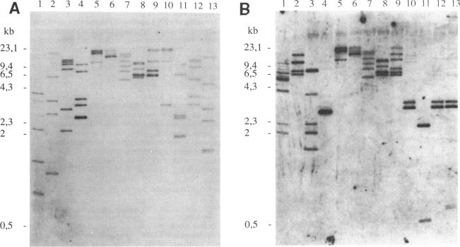
The circular physical map of the pathogenic bacterium Listeria monocytogenes LO28 (serovar 1/2c) was established by using pulsed-field gel electrophoresis. The L. monocytogenes chromosome contains eight NotI fragments of 1,100, 940, 400, 335, 280, 45, 30, and 20 kb in size and eight Sse8387I fragments of 860, 680, 680, 370, 335, 130, 70, and 25 kb. Therefore, the total length of the genome is 3,150 kb. To order the NotI fragments on the chromosome, we used a strategy which can be of general use. We first cloned chromosomal HindIII or EcoRI fragments in pBR322. DNA extracted from the total libraries was digested by NotI and ligated to a NotI-kanamycin resistance cassette obtained by cutting Tn5 with NotI. After transformation in Escherichia coli, kanamycin-resistant clones originating from NotI-containing EcoRI or HindIII fragments were isolated. The two EcoRI-NotI or HindIII-NotI fragments of each recombinant plasmid were isolated and used as probes on Southern blot hybridizations to identify and link the corresponding NotI fragments. Seven NotI fragments were ordered in this way. The last junction was demonstrated by partial digest analysis. All L. monocytogenes genes identified so far as well as the six rRNA operons were localized on the NotI map. Regions homologous to genes from closely related bacteria were also detected and localized. Southern blot analysis of simple Sse8387I digests or double Sse8387I-NotI digests probed with the various NotI probes allowed us to align the Sse8387I fragments and localize the single SfiI site, resulting in the establishment of the first genetic and physical map of the L. monocytogenes chromosome.
通过脉冲场凝胶电泳建立了致病性单核细胞增生李斯特菌LO28(血清型1/2c)的环状物理图谱。单核细胞增生李斯特菌染色体包含8个大小分别为1100、940、400、335、280、45、30和20 kb的NotI片段以及8个大小分别为860、680、680、370、335、130、70和25 kb的Sse8387I片段。因此,基因组的总长度为3150 kb。为了在染色体上排列NotI片段,我们采用了一种通用策略。我们首先将染色体HindIII或EcoRI片段克隆到pBR322中。从总文库中提取的DNA用NotI消化,并与通过用NotI切割Tn5获得的NotI - 卡那霉素抗性盒连接。在大肠杆菌中转化后,分离出来自含NotI的EcoRI或HindIII片段的卡那霉素抗性克隆。分离每个重组质粒的两个EcoRI - NotI或HindIII - NotI片段,并将其用作Southern印迹杂交的探针,以鉴定和连接相应的NotI片段。通过这种方式排列了7个NotI片段。最后一个连接点通过部分消化分析得到证实。到目前为止鉴定出的所有单核细胞增生李斯特菌基因以及6个rRNA操纵子都定位在NotI图谱上。还检测并定位了与密切相关细菌基因同源的区域。用各种NotI探针探测简单的Sse8387I消化产物或双重Sse8387I - NotI消化产物的Southern印迹分析使我们能够排列Sse8387I片段并定位单个SfiI位点,从而建立了单核细胞增生李斯特菌染色体的首张遗传和物理图谱。