Nistal M, Santamaría L, Paniagua R
Department of Morphology, School of Medicine, Autonomous University, Madrid, Spain.
J Anat. 1992 Feb;180 ( Pt 1)(Pt 1):97-104.
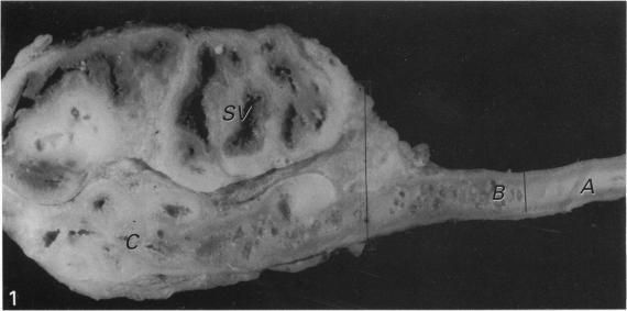
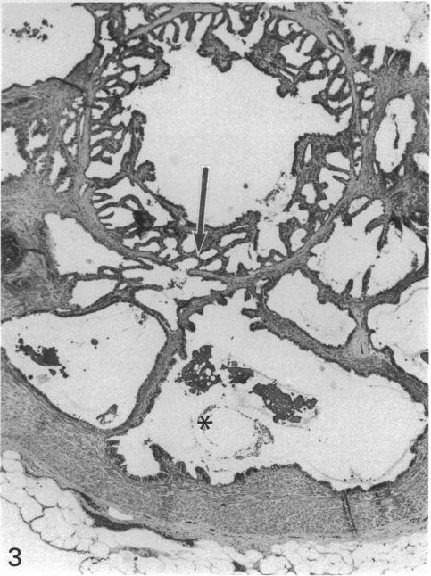
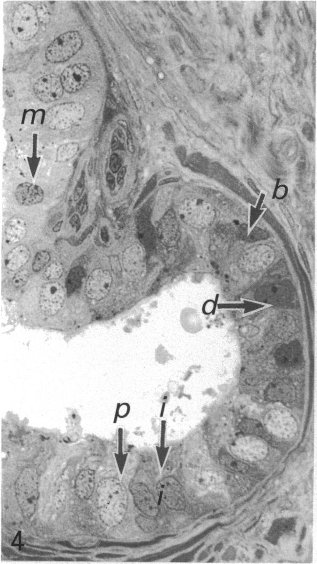
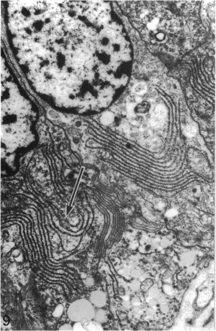
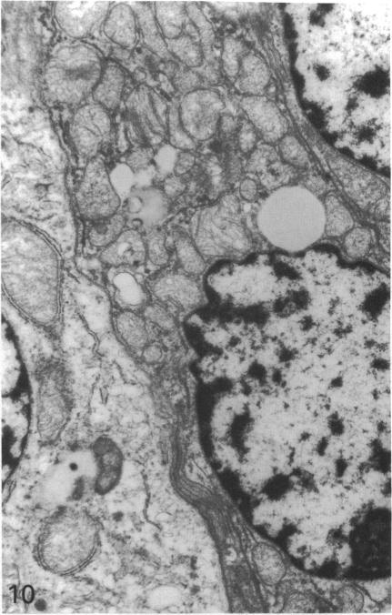
In order to compare the histology of the ampulla of the ductus deferens with that of the other segments of the duct in man, the seminal vesicle and the adjacent 13-15 cm of the ductus deferens were obtained during cystectomy from 15 adult men, and were processed for light and electron microscopy. Each ductus deferens specimen was divided into 3 segments: segment A or initial segment (the most proximal to the testis) showing a smooth outer surface and, on section, a uniform lumen and absence of mucosal invaginations; segment B (1.5-4 cm) showing a smooth outer surface and, on section, small cavities in the mucosa; and segment C or ampulla (3-4 cm), which was easily recognisable because of the cerebriform pattern on its outer surface. Segment A showed the usual histological pattern reported in studies of the human ductus deferens. Segment B consisted of mucosa, muscularis mucosae, submucosa, muscular coat and adventitia. The epithelial lining formed multiple branched invaginations in the lamina propria and submucosa giving rise to glandular structures. The lumen of the duct and the glands were lined by the same cell types: (1) basal cells; (2) mitochondrion-rich cells; and (3) columnar cells with the ultrastructural features of glycoprotein-secreting cells. The latter cells could be classified into 3 subtypes suggesting different stages of development: (a) with abundant mitochondria; (b) with abundant rough endoplasmic reticulum; and (c) with abundant secretory granules. Segment C or the ampulla showed the same histology as segment B except for the presence of many diverticula in the ampulla.(ABSTRACT TRUNCATED AT 250 WORDS)
为了比较人类输精管壶腹部与输精管其他节段、精囊及输精管相邻13 - 15厘米段的组织学,在15例成年男性膀胱切除术中获取了精囊及输精管相邻段,并进行光镜和电镜处理。每个输精管标本分为3段:A段或起始段(最靠近睾丸),外表面光滑,切片显示管腔均匀且无黏膜内陷;B段(1.5 - 4厘米),外表面光滑,切片显示黏膜中有小腔;C段或壶腹部(3 - 4厘米),因其外表面呈脑回状图案而易于辨认。A段呈现出人类输精管研究中报道的常见组织学模式。B段由黏膜、黏膜肌层、黏膜下层、肌层和外膜组成。上皮衬里在固有层和黏膜下层形成多个分支内陷,产生腺结构。输精管腔和腺体由相同类型的细胞内衬:(1) 基底细胞;(2) 富含线粒体的细胞;(3) 具有糖蛋白分泌细胞超微结构特征的柱状细胞。后一种细胞可分为3个亚型,提示不同的发育阶段:(a) 线粒体丰富;(b) 粗面内质网丰富;(c) 分泌颗粒丰富。C段或壶腹部除壶腹部存在许多憩室外,组织学与B段相同。(摘要截断于250字)