Kuefer R, Hofer M D, Altug V, Zorn C, Genze F, Kunzi-Rapp K, Hautmann R E, Gschwend J E
1Department of Urology, University of Ulm, Prittwitz-Strasse 43, 89075 Ulm, Germany.
Br J Cancer. 2004 Jan 26;90(2):535-41. doi: 10.1038/sj.bjc.6601510.
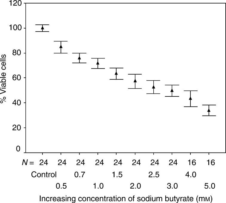
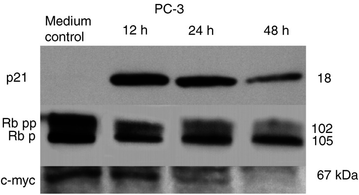
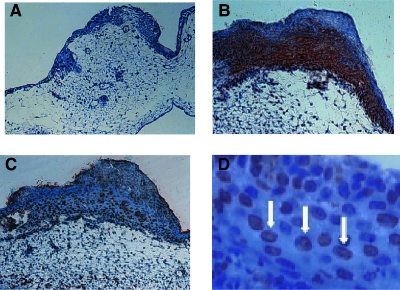
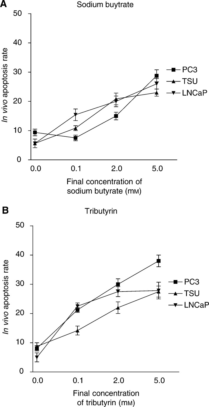
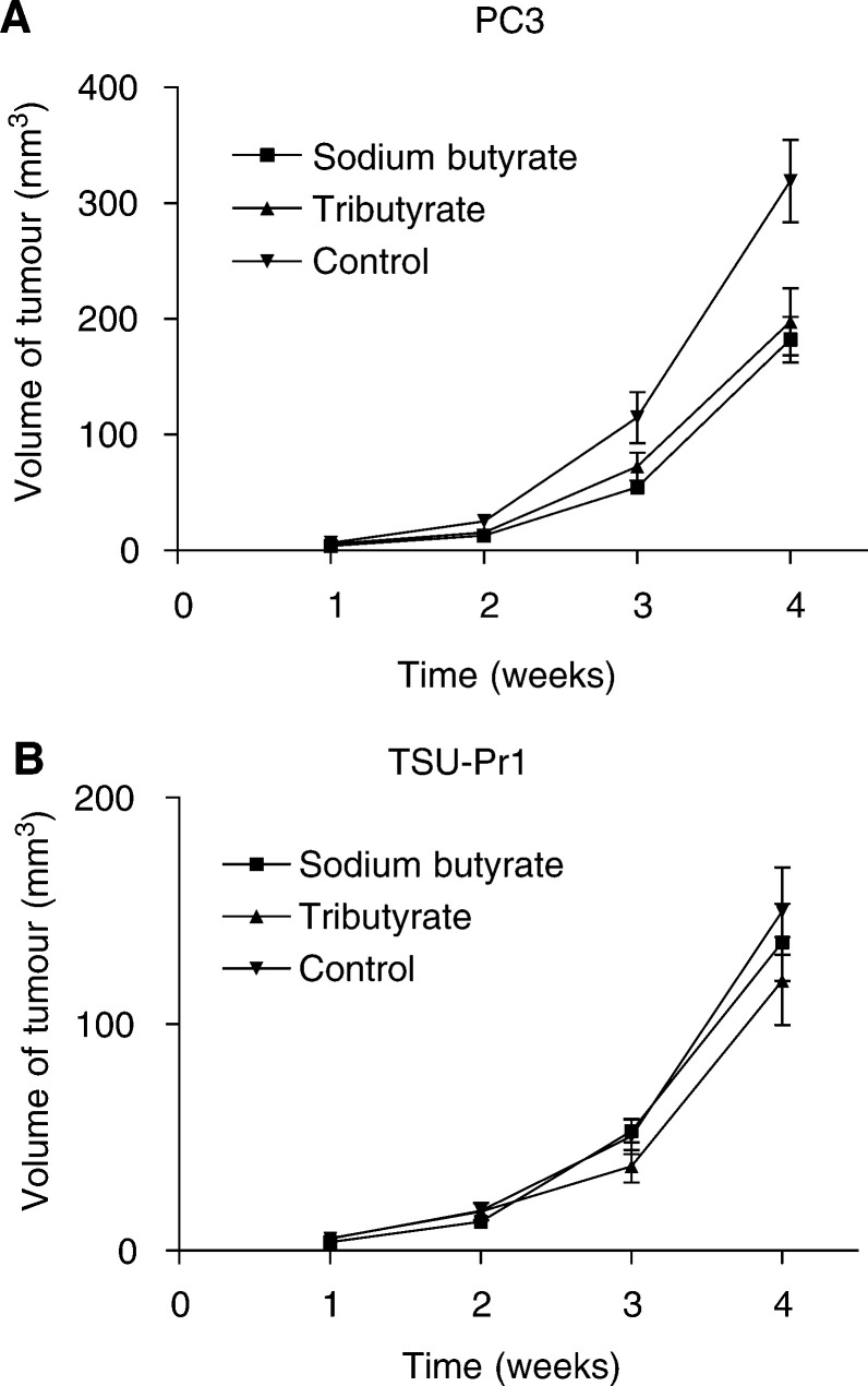
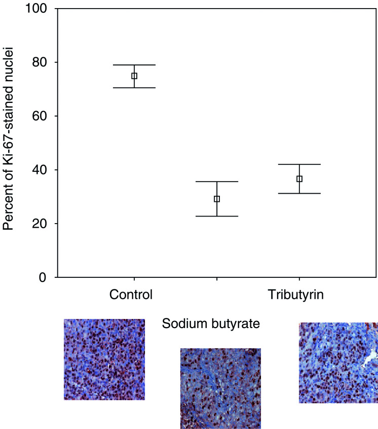
Histone deacetylase inhibitors (HDACs) are known to exhibit antiproliferative effects on various carcinoma cells. In this study, the in vivo efficiency of two HDACs, sodium butyrate and tributyrin, on prostate cancer growth inhibition were investigated. To gain an insight into the possible underlying pathways, cell culture experiments were performed focusing on the expression of p21, Rb and c-myc. For in vivo testing, prostate cancer cell lines (PC3 and TSU-Pr1) were seeded on the chorioallantois membrane (CAM) and implanted in a xenograft model using nude mice. Standard Western blot analysis was performed for protein expression of p21, Rb and c-myc in HDAC-treated vs untreated prostate cancer cells. Both sodium butyrate and tributyrin had a considerable treatment effect on microtumours on the chicken egg at already very low concentrations of 0.1 mM. Tributyrin-treated tumours showed the strongest effect with 38% apoptotic nuclei in the prostate cancer cell line PC3. In the mouse model, there was almost no difference between sodium butyrate and tributyrin. In untreated animals the tumours were almost double the size 4 weeks after implantation. Tumours of the treatment groups had a significantly lower percentage of Ki-67-positive-stained nuclei. As demonstrated by Western blot analysis, these effects seem to be independent of p53 status and a pathway via p21-Rb-c-myc is possibly involved. In this study we have demonstrated a substantial in vivo treatment effect, which can be induced by the application of sodium butyrate or the orally applicable tributyrin in human prostate cancer. The given results may provide the rationale to apply these drugs in well-controlled clinical trials in patients being at high risk of recurrence after specific therapy or in patients with locally or distant advanced prostate cancer.
组蛋白去乙酰化酶抑制剂(HDACs)已知对多种癌细胞具有抗增殖作用。在本研究中,研究了两种HDACs(丁酸钠和三丁酸甘油酯)对前列腺癌生长抑制的体内效果。为深入了解可能的潜在途径,进行了细胞培养实验,重点关注p21、Rb和c-myc的表达。为进行体内测试,将前列腺癌细胞系(PC3和TSU-Pr1)接种于鸡胚绒毛尿囊膜(CAM)上,并植入裸鼠异种移植模型中。对HDAC处理组和未处理组的前列腺癌细胞进行标准蛋白质免疫印迹分析,以检测p21、Rb和c-myc的蛋白质表达。丁酸钠和三丁酸甘油酯在极低浓度(0.1 mM)时对鸡蛋上的微肿瘤均有显著的治疗效果。三丁酸甘油酯处理的肿瘤效果最强,前列腺癌细胞系PC3中有38%的凋亡细胞核。在小鼠模型中,丁酸钠和三丁酸甘油酯之间几乎没有差异。在未处理的动物中,植入后4周肿瘤大小几乎翻倍。治疗组肿瘤中Ki-67阳性染色细胞核的百分比显著较低。蛋白质免疫印迹分析表明,这些作用似乎与p53状态无关,可能涉及一条通过p21-Rb-c-myc的途径。在本研究中,我们证明了丁酸钠或口服可用的三丁酸甘油酯对人前列腺癌具有显著的体内治疗效果。给出的结果可能为在特定治疗后复发风险高的患者或局部或远处晚期前列腺癌患者中,在严格控制的临床试验中应用这些药物提供理论依据。