Sakakura C, Takemura M, Hagiwara A, Shimomura K, Miyagawa K, Nakashima S, Yoshikawa T, Takagi T, Kin S, Nakase Y, Fujiyama J, Hayasizaki Y, Okazaki Y, Yamagishi H
Department of Digestive Surgery, Kyoto Prefectural University of Medicine, Kamigyo-ku, Kawaramachi-dori, Kyoto 602-8566, Japan.
Br J Cancer. 2004 Feb 9;90(3):665-71. doi: 10.1038/sj.bjc.6601544.
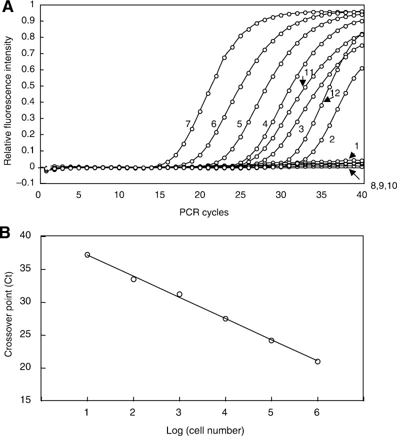
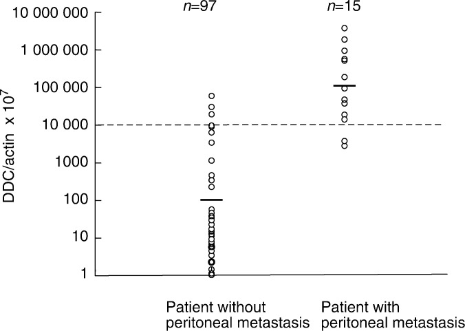
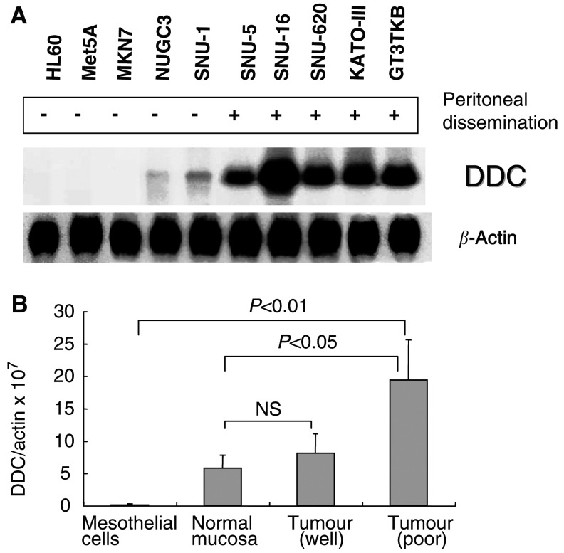
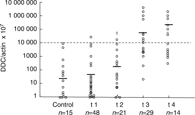
We previously performed a global analysis of the gene expression of gastric cancer cell lines established from metastases to the peritoneal cavity with the cDNA microarray method, which made it possible to analyse the expression of approximately 21168 genes for the identification of novel markers for the detection of micrometastases in the peritoneal cavity. One of the upregulated genes is dopa decarboxylase (DDC), which is responsible for the synthesis of the key neurotransmitters dopamine and serotonine. We have examined its potential as a novel marker for the detection of peritoneal micrometastases of gastric cancer.DDC mRNA in the peritoneal wash from 112 gastric cancer patients was quantified for comparison of carcinoembryonic antigen (CEA) mRNA by means of real-time reverse transcriptase-polymerase chain reaction (RT-PCR) with a fluorescently labelled probe to predict peritoneal recurrence. The quantity of DDC and CEA correlated with wall penetration. Real-time RT-PCR could quantitate 10-10(6) DDC-expressing gastric cancer cells per 10(7) mesothelial cells. The cutoff value was set at the upper limit of the quantitative value for noncancer patients, and those above this cutoff value constituted the micrometastasis (MM+) group. Of 15 cases with peritoneal dissemination, 13 were MM+DDC (87% sensitivity), and one of 48 t1 cases was MM+ (98% specificity). DDC levels in peritoneal washes from patients with synchronous peritoneal metastases were more than 50 times higher than in those from patients without metastasis (P<0.01). For 15 cases of peritoneal dissemination (seven cases were cytologically positive), DDC was positive in 13 cases (87% sensitivity), but CEA failed to detect micrometastases in four cases (73% sensitivity), indicating that DDC is in some cases superior to CEA for the detection of peritoneal micrometastases of gastric cancer in terms of sensitivity as well as specificity, especially for poorly differentiated adenocarcinomas. A combination of CEA and DDC improved the accuracy of diagnosis up to 94%. These results suggest that DDC is potentially a novel marker for peritoneal dissemination of gastric cancer and that quantitative RT-PCR of DDC is reliable and efficient for the selection of patients for adjuvant intraperitoneal chemotherapy to prevent peritoneal recurrence.
我们之前使用cDNA微阵列方法对从转移至腹腔的胃癌细胞系进行了基因表达的全面分析,这使得分析约21168个基因的表达成为可能,以鉴定用于检测腹腔微转移的新型标志物。上调基因之一是多巴脱羧酶(DDC),它负责合成关键神经递质多巴胺和血清素。我们研究了其作为检测胃癌腹腔微转移新型标志物的潜力。通过实时逆转录聚合酶链反应(RT-PCR)和荧光标记探针,对112例胃癌患者腹腔灌洗液中的DDC mRNA进行定量,以比较癌胚抗原(CEA)mRNA,从而预测腹腔复发。DDC和CEA的量与壁层浸润相关。实时RT-PCR能够定量每10⁷个间皮细胞中表达DDC的10⁻¹⁰⁶个胃癌细胞。将临界值设定为非癌症患者定量值的上限,高于此临界值的患者构成微转移(MM+)组。在15例有腹腔播散的病例中,13例为MM+DDC(敏感性87%),48例t1期病例中有1例为MM+(特异性98%)。同步发生腹腔转移患者腹腔灌洗液中的DDC水平比无转移患者高50倍以上(P<0.01)。对于15例腹腔播散病例(7例细胞学阳性),DDC在13例中呈阳性(敏感性87%),但CEA在4例中未能检测到微转移(敏感性73%),这表明在检测胃癌腹腔微转移方面,DDC在敏感性和特异性方面在某些情况下优于CEA,尤其是对于低分化腺癌。CEA和DDC联合使用可将诊断准确性提高至94%。这些结果表明,DDC可能是胃癌腹腔播散的新型标志物,并且DDC的定量RT-PCR对于选择接受辅助性腹腔内化疗以预防腹腔复发的患者是可靠且有效的。