Thordarson Gudmundur, Slusher Nicole, Leong Harriet, Ochoa Dafne, Rajkumar Lakshmanaswamy, Guzman Raphael, Nandi Satyabrata, Talamantes Frank
Department of Molecular, Cell, and Developmental Biology, University of California, Santa Cruz, California, USA.
Breast Cancer Res. 2004;6(4):R423-36. doi: 10.1186/bcr812. Epub 2004 Jun 4.
Pregnancy protects against breast cancer development in humans and rats. Parous rats have persistently reduced circulating levels of growth hormone, which may affect the activity of the growth hormone/insulin-like growth factor (IGF)-I axis. We investigated the effects of IGF-I on parity-associated protection against mammary cancer.
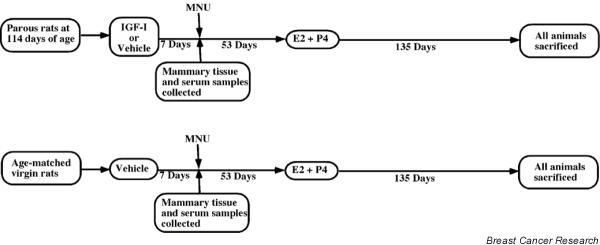
Three groups of rats were evaluated in the present study: IGF-I-treated parous rats; parous rats that did not receive IGF-I treatment; and age-matched virgin animals, which also did not receive IGF-I treatment. Approximately 60 days after N-methyl-N-nitrosourea injection, IGF-I treatment was discontinued and all of the animal groups were implanted with a silastic capsule containing 17beta-estradiol and progesterone. The 17beta-estradiol plus progesterone treatment continued for 135 days, after which the animals were killed.
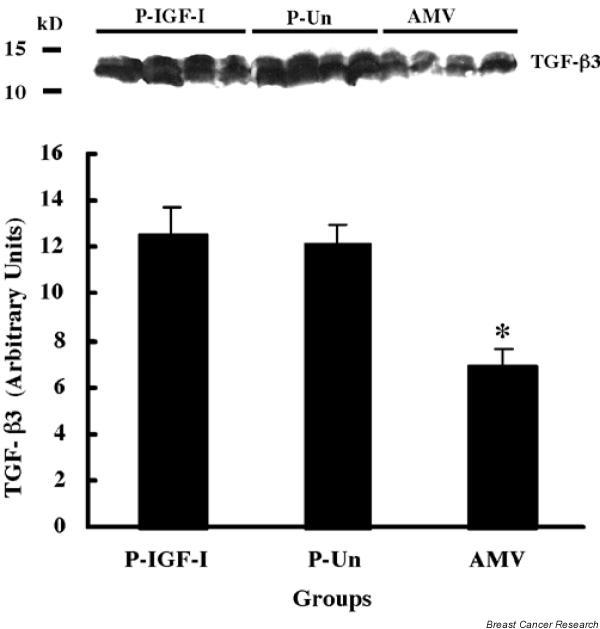
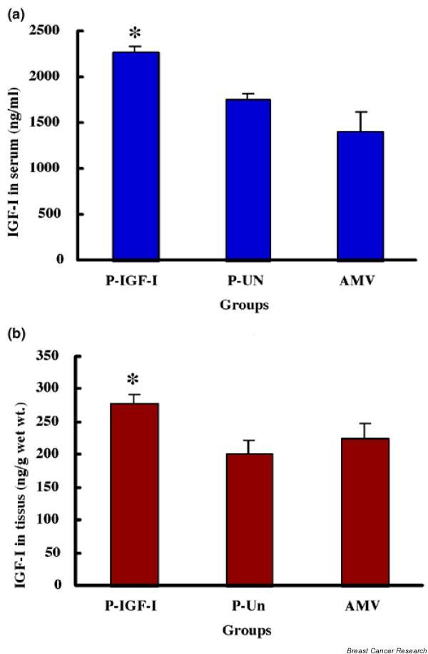
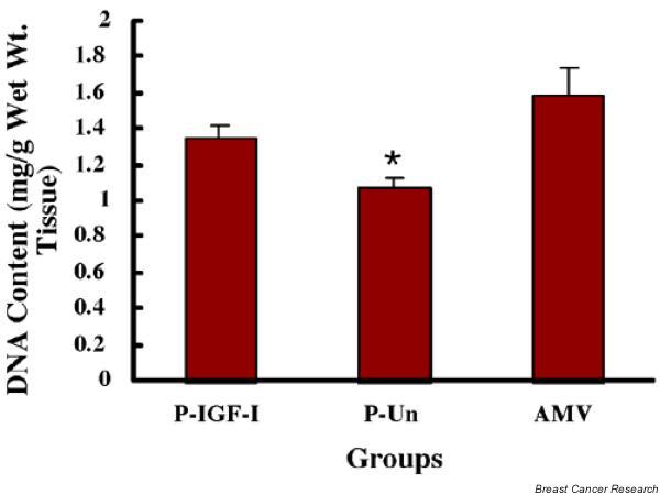
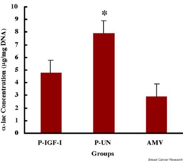
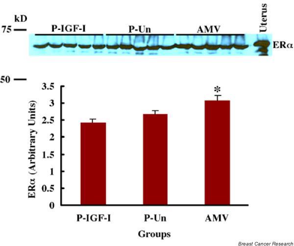
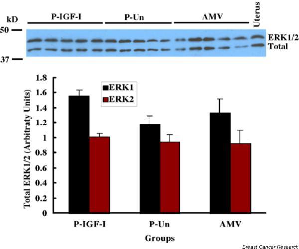
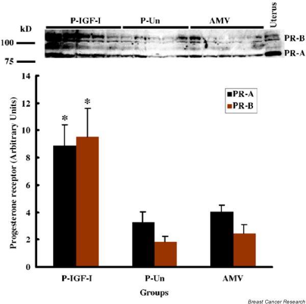
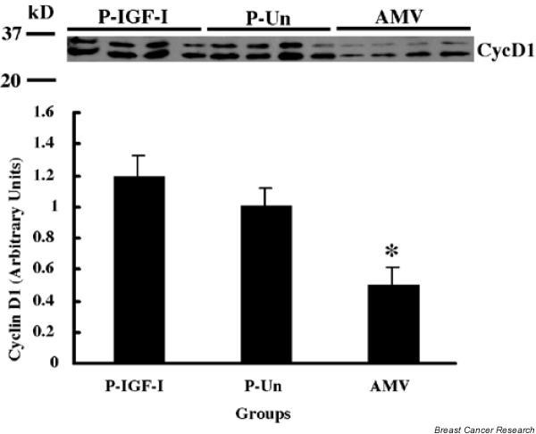
IGF-I treatment of parous rats increased mammary tumor incidence to 83%, as compared with 16% in parous rats treated with 17beta-estradiol plus progesterone only. Tumor incidence and average number of tumors per animal did not differ between IGF-I-treated parous rats and age-matched virgin rats. At the time of N-methyl-N-nitrosourea exposure, DNA content was lowest but the alpha-lactalbumin concentration highest in the mammary glands of untreated parous rats in comparison with age-matched virgin and IGF-I-treated parous rats. The protein levels of estrogen receptor-alpha in the mammary gland was significantly higher in the age-matched virgin animals than in untreated parous and IGF-I-treated parous rats. Phosphorylation (activation) of the extracellular signal-regulated kinase-1/2 (ERK1/2) and expression of the progesterone receptor were both increased in IGF-I-treated parous rats, as compared with those in untreated parous and age-matched virgin rats. Expressions of cyclin D1 and transforming growth factor-beta3 in the mammary gland were lower in the age-matched virgin rats than in the untreated parous and IGF-I-treated parous rats.
We argue that tumor initiation (transformation and fixation of mutations) may be similar in parous and age-matched virgin animals, suggesting that the main differences in tumor formation lie in differences in tumor progression caused by the altered hormonal environment associated with parity. Furthermore, we provide evidence supporting the notion that tumor growth promotion seen in IGF-I-treated parous rats is caused by activation of estrogen receptor-alpha via the Raf/Ras/mitogen-activated protein kinase cascade.
怀孕可预防人类和大鼠患乳腺癌。经产大鼠的生长激素循环水平持续降低,这可能会影响生长激素/胰岛素样生长因子(IGF)-I轴的活性。我们研究了IGF-I对经产相关的乳腺癌预防作用的影响。
本研究评估了三组大鼠:接受IGF-I治疗的经产大鼠;未接受IGF-I治疗的经产大鼠;以及年龄匹配的未接受IGF-I治疗的处女鼠。在注射N-甲基-N-亚硝基脲后约60天,停止IGF-I治疗,所有动物组均植入含有17β-雌二醇和孕酮的硅橡胶胶囊。17β-雌二醇加孕酮治疗持续135天,之后处死动物。
与仅接受17β-雌二醇加孕酮治疗的经产大鼠中16%的乳腺肿瘤发生率相比,接受IGF-I治疗的经产大鼠的乳腺肿瘤发生率增加到83%。接受IGF-I治疗的经产大鼠与年龄匹配的处女鼠之间的肿瘤发生率和每只动物的平均肿瘤数量没有差异。在接触N-甲基-N-亚硝基脲时,与年龄匹配的处女鼠和接受IGF-I治疗的经产大鼠相比,未治疗的经产大鼠乳腺中的DNA含量最低,但α-乳白蛋白浓度最高。年龄匹配的处女鼠乳腺中雌激素受体-α的蛋白水平显著高于未治疗的经产大鼠和接受IGF-I治疗的经产大鼠。与未治疗的经产大鼠和年龄匹配的处女鼠相比,接受IGF-I治疗的经产大鼠中细胞外信号调节激酶-1/2(ERK1/2)的磷酸化(激活)和孕酮受体的表达均增加。年龄匹配的处女鼠乳腺中细胞周期蛋白D1和转化生长因子-β3的表达低于未治疗的经产大鼠和接受IGF-I治疗的经产大鼠。
我们认为经产大鼠和年龄匹配的处女鼠在肿瘤起始(突变的转化和固定)方面可能相似,这表明肿瘤形成的主要差异在于与经产相关的激素环境改变所导致的肿瘤进展差异。此外,我们提供的证据支持以下观点:在接受IGF-I治疗的经产大鼠中观察到的肿瘤生长促进是由雌激素受体-α通过Raf/Ras/丝裂原活化蛋白激酶级联反应激活所致。