Choesmel Valérie, Pierga Jean-Yves, Nos Claude, Vincent-Salomon Anne, Sigal-Zafrani Brigitte, Thiery Jean-Paul, Blin Nathalie
UMR144 CNRS, Research Division, Institut Curie, Paris, France.
Breast Cancer Res. 2004;6(5):R556-70. doi: 10.1186/bcr898. Epub 2004 Jul 29.
Improving technologies for the detection and purification of bone marrow (BM) micrometastatic cells in breast cancer patients should lead to earlier prognosis of the risk of relapse and should make it possible to design more appropriate therapies. The technique used has to overcome the challenges resulting from the small number of target cells (one per million hematopoietic cells) and the heterogeneous expression of micrometastatic cell markers. In the present study, we have assessed the clinical relevance of current methods aimed at detecting rare disseminated carcinoma cells.
BM aspirates from 32 carcinoma patients were screened for the presence of micrometastatic cells positive for epithelial cell adhesion molecule and positive for cytokeratins, using optimized immunodetection methods. A comparison with data obtained for 46 control BM aspirates and a correlation with the clinical status of patients were performed.
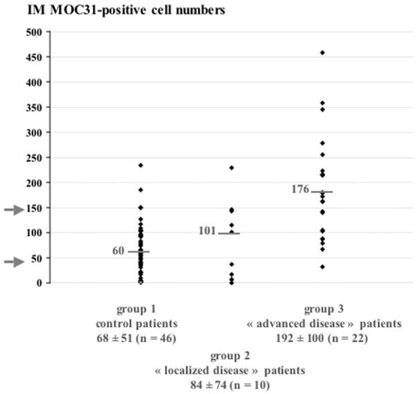
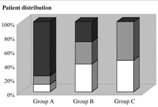
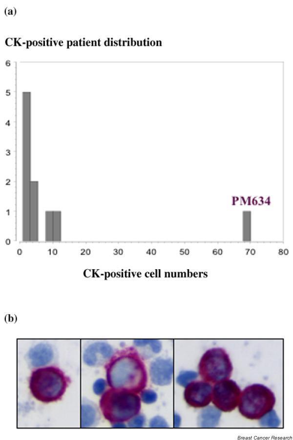
We developed a sensitive and efficient immunomagnetic protocol for the enrichment of BM micrometastases. This method was used to divide 32 breast carcinoma patients into three categories according to their epithelial cell adhesion molecule status. These categories were highly correlated with the recently revised American Joint Committee on Cancer staging system for breast cancer, demonstrating the clinical relevance of this simple and reliable immunomagnetic technique. We also evaluated immunocytochemical detection of cytokeratin-positive cells and cytomorphological parameters. Immunocytochemistry-based methods for the detection of BM micrometastases did not provide any information about the clinical status of patients, but helped to refine the immunomagnetic data by confirming the presence of micrometastases in some cases. We also tested a new density gradient centrifugation system, able to enrich the tumor fraction of BM specimens by twofold to threefold as compared with standard Ficoll methods.
These improved methods for the detection of micrometastatic cells in patient BM should help clinicians to predict the clinical status of breast cancer patients at the time of surgery or treatment.
改进乳腺癌患者骨髓(BM)微转移细胞检测和纯化技术应能更早地预测复发风险,并使设计更合适的治疗方案成为可能。所采用的技术必须克服因靶细胞数量少(每百万造血细胞中一个)以及微转移细胞标志物表达异质性所带来的挑战。在本研究中,我们评估了旨在检测罕见播散癌细胞的现有方法的临床相关性。
使用优化的免疫检测方法,对32例癌症患者的骨髓抽吸物进行筛查,以检测上皮细胞粘附分子阳性且细胞角蛋白阳性的微转移细胞。将其与46例对照骨髓抽吸物的数据进行比较,并与患者的临床状态进行相关性分析。
我们开发了一种灵敏且高效的免疫磁选方案用于富集骨髓微转移灶。该方法用于根据上皮细胞粘附分子状态将32例乳腺癌患者分为三类。这些类别与最近修订的美国癌症联合委员会乳腺癌分期系统高度相关,证明了这种简单可靠的免疫磁选技术的临床相关性。我们还评估了细胞角蛋白阳性细胞的免疫细胞化学检测和细胞形态学参数。基于免疫细胞化学的骨髓微转移检测方法未提供任何有关患者临床状态的信息,但通过在某些情况下确认微转移灶的存在,有助于完善免疫磁选数据。我们还测试了一种新的密度梯度离心系统,与标准Ficoll方法相比,该系统能够将骨髓标本的肿瘤部分富集两到三倍。
这些改进的检测患者骨髓中微转移细胞的方法应有助于临床医生在手术或治疗时预测乳腺癌患者的临床状态。