Williams Anthony, Chung Jaebum, Ou Xiaoze, Zheng Guoan, Rawal Siddarth, Ao Zheng, Datar Ram, Yang Changhuei, Cote Richard
University of Miami, Miller School of Medicine, Department of Pathology, 1501 NW 10th Avenue BRB 742, Miami, Florida 33136bUniversity of Miami, Dr. John T. Macdonald Foundation Biomedical Nanotechnology Institute (BioNIUM), 1501 NW 10th Avenue BRB 714, Mi.
California Institute of Technology, Departments of Electrical Engineering, Bioengineering, and Medical Engineering, 1200 East California Boulevard MC 136-93, Pasadena, California 91125.
J Biomed Opt. 2014 Jun;19(6):066007. doi: 10.1117/1.JBO.19.6.066007.
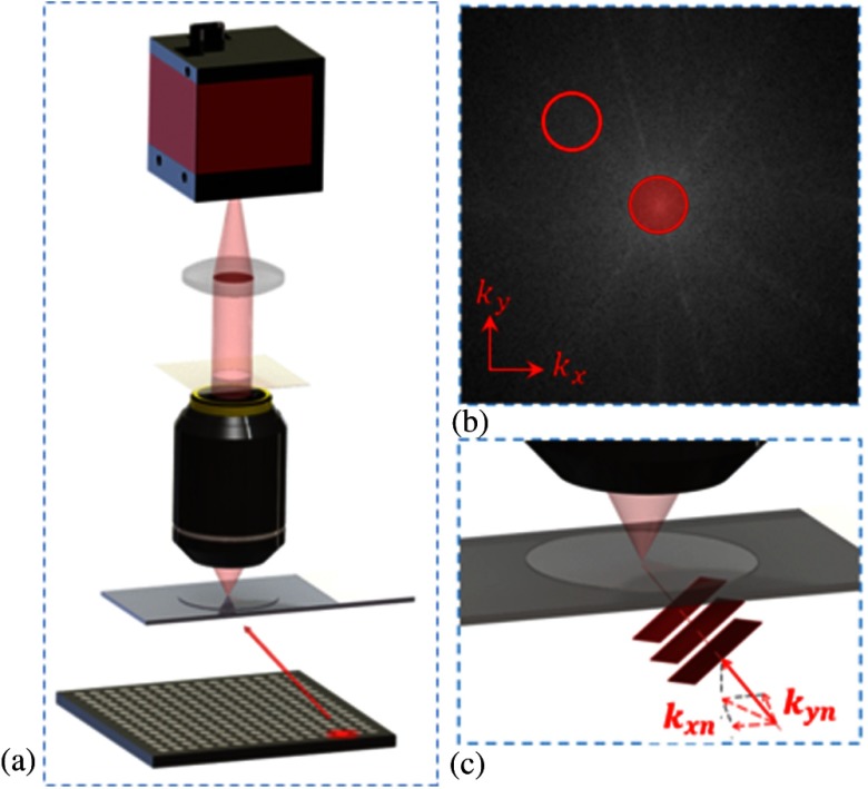
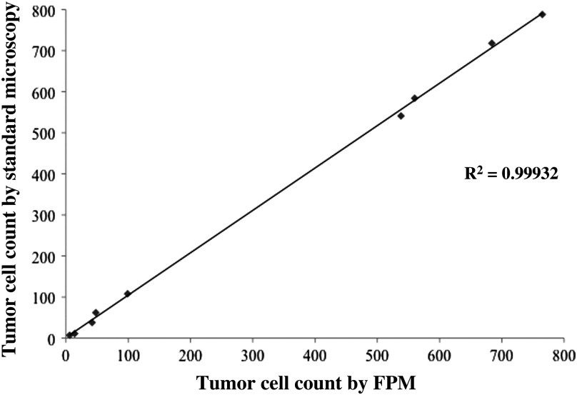
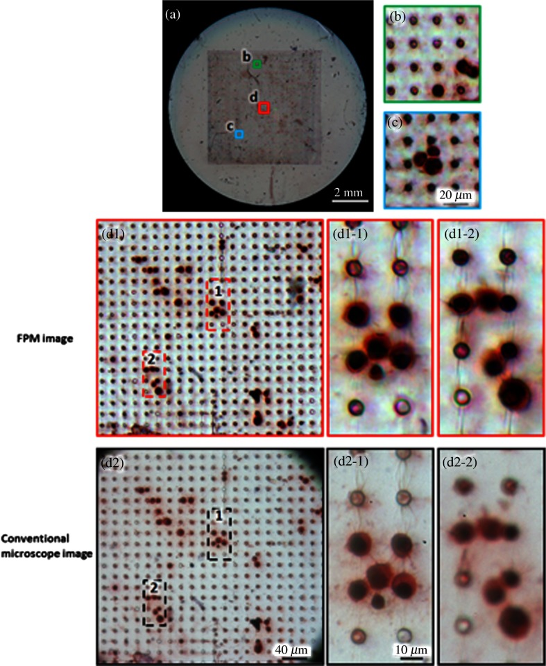
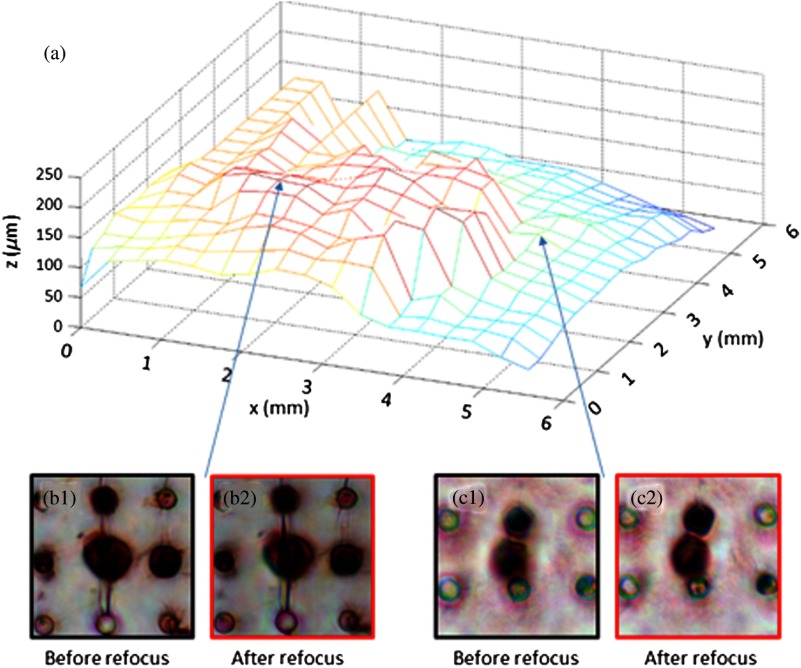
Circulating tumor cells (CTCs) are recognized as a candidate biomarker with strong prognostic and predictive potential in metastatic disease. Filtration-based enrichment technologies have been used for CTC characterization, and our group has previously developed a membrane microfilter device that demonstrates efficacy in model systems and clinical blood samples. However, uneven filtration surfaces make the use of standard microscopic techniques a difficult task, limiting the performance of automated imaging using commercially available technologies. Here, we report the use of Fourier ptychographic microscopy (FPM) to tackle this challenge. Employing this method, we were able to obtain high-resolution color images, including amplitude and phase, of the microfilter samples over large areas. FPM's ability to perform digital refocusing on complex images is particularly useful in this setting as, in contrast to other imaging platforms, we can focus samples on multiple focal planes within the same frame despite surface unevenness. In model systems, FPM demonstrates high image quality, efficiency, and consistency in detection of tumor cells when comparing corresponding microfilter samples to standard microscopy with high correlation (R² = 0.99932). Based on these results, we believe that FPM will have important implications for improved, high throughput, filtration-based CTC analysis, and, more generally, image analysis of uneven surfaces.
循环肿瘤细胞(CTCs)被认为是一种在转移性疾病中具有强大预后和预测潜力的候选生物标志物。基于过滤的富集技术已用于CTCs表征,我们小组此前开发了一种膜微滤装置,该装置在模型系统和临床血样中显示出有效性。然而,过滤表面不均匀使得使用标准显微镜技术成为一项艰巨任务,限制了使用市售技术进行自动成像的性能。在此,我们报告使用傅里叶叠层显微镜(FPM)来应对这一挑战。采用这种方法,我们能够在大面积上获得微滤样品的高分辨率彩色图像,包括幅度和相位。FPM对复杂图像进行数字重聚焦的能力在这种情况下特别有用,因为与其他成像平台相比,尽管表面不均匀,我们仍可以在同一帧内对多个焦平面上的样品进行聚焦。在模型系统中,将相应的微滤样品与标准显微镜进行比较时,FPM在检测肿瘤细胞方面表现出高图像质量、效率和一致性,相关性很高(R² = 0.99932)。基于这些结果,我们认为FPM将对改进的、高通量的基于过滤的CTCs分析以及更一般的不均匀表面图像分析具有重要意义。