Rossi Michele, Cantisani Vito, Salvatori Filippo Maria, Rebonato Alberto, Greco Laura, Giglio Luigi, Guido Giampiero, Pagliara Elisa, David Vincenzo
Department of Radiology, "S, Andrea" Hospital-II Faculty "La Sapienza" University, Rome,00100, Italy.
BMC Med Imaging. 2004 Aug 25;4(1):3. doi: 10.1186/1471-2342-4-3.
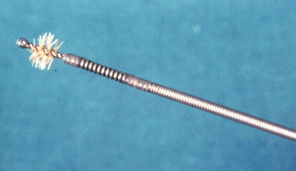
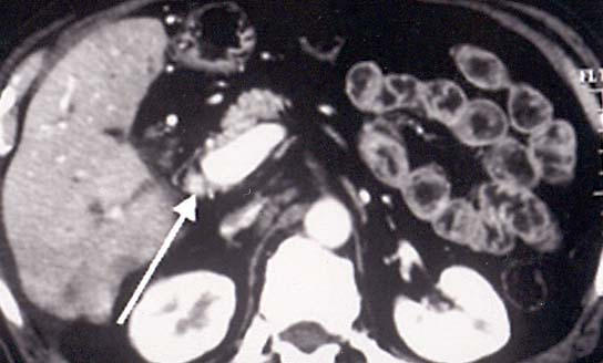
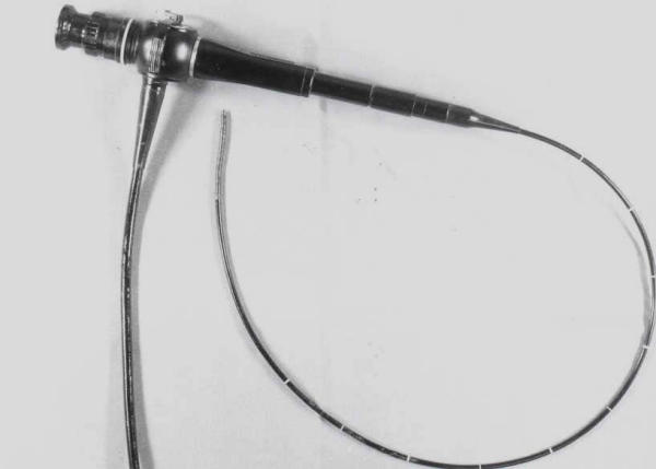
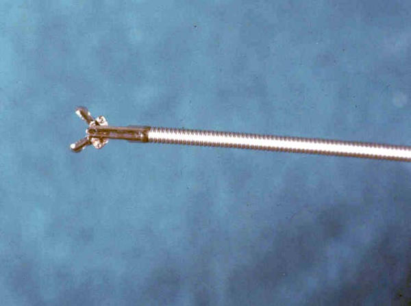
Despite the sophisticated cross sectional image techniques currently available, a number of biliary stenosis or obstructions remain of an uncertain nature. In these pathological conditions, an "intrinsic" parietal alteration is the cause of biliary obstruction and it is very difficult to differentiate benign from malignant lesions using cross-sectional imaging procedures alone. We evaluated the efficacy of different endoluminal techniques to achieve a definitive pathological diagnosis in these situations. METHODS: Eighty patients underwent brushing, and or biopsy of the biliary tree through an existing transhepatic biliary drainage route. A subcoort of 12 patients needed balloon-dilatation of the bile duct and the material covering the balloon surface was also sent for pathological examination (balloon surface sampling). Pathological results were compared with surgical findings or with long-term clinical and instrumental follow-ups. Success rates, sensitivity, specificity, accuracy, confidential intervals, positive predictive value and negative predictive value of the three percutaneous techniques in differentiating benign from malignant disease were assessed.The agreement coefficient of biopsy and brushing with final diagnosis was calculated using the Cohen's "K" value. RESULTS: Fifty-six patients had malignant strictures confirmed by surgery, histology, and by clinical follow-ups. Success rates of brushing, balloon surface sampling, and biopsy were 90.7, 100, and 100%, respectively. The comparative efficacy of brushing, balloon-surface sampling, and biopsy resulted as follows: sensitivity of 47.8, 87.5, and 92.1%, respectively; specificity of 100% for all the techniques; accuracy of 69.2, 91.7 and 93.6%, Positive Predictive Value of 100% for all the procedures and Negative Predictive Value of 55, 80, and 75%, respectively. CONCLUSIONS: Percutaneous endoluminal biopsy is more accurate and sensitive than percutaneous bile duct brushing in the detection of malignant diseases (p < 0.01).
尽管目前有先进的横断面成像技术,但仍有一些胆管狭窄或梗阻的性质不确定。在这些病理情况下,“内在的”壁层改变是胆管梗阻的原因,仅使用横断面成像程序很难区分良性和恶性病变。我们评估了不同腔内技术在这些情况下获得明确病理诊断的疗效。
80例患者通过现有的经肝胆汁引流途径对胆管进行刷检和/或活检。12例患者的亚组需要对胆管进行球囊扩张,球囊表面覆盖的材料也送去进行病理检查(球囊表面采样)。将病理结果与手术结果或长期临床及影像学随访结果进行比较。评估了三种经皮技术在区分良性和恶性疾病方面的成功率、敏感性、特异性、准确性、可信区间、阳性预测值和阴性预测值。使用科恩“K”值计算活检和刷检与最终诊断的一致性系数。
56例患者经手术、组织学和临床随访证实为恶性狭窄。刷检、球囊表面采样和活检的成功率分别为90.7%、100%和100%。刷检、球囊表面采样和活检的比较疗效如下:敏感性分别为47.8%、87.5%和92.1%;所有技术的特异性均为100%;准确性分别为69.2%、91.7%和93.6%,所有程序的阳性预测值均为100%,阴性预测值分别为55%、80%和75%。
在检测恶性疾病方面,经皮腔内活检比经皮胆管刷检更准确、更敏感(p<0.01)。