Thomas J L, Feder T J, Webb W W
Department of Physics, Cornell University, Ithaca, New York 14853.
Biophys J. 1992 May;61(5):1402-12. doi: 10.1016/S0006-3495(92)81946-X.
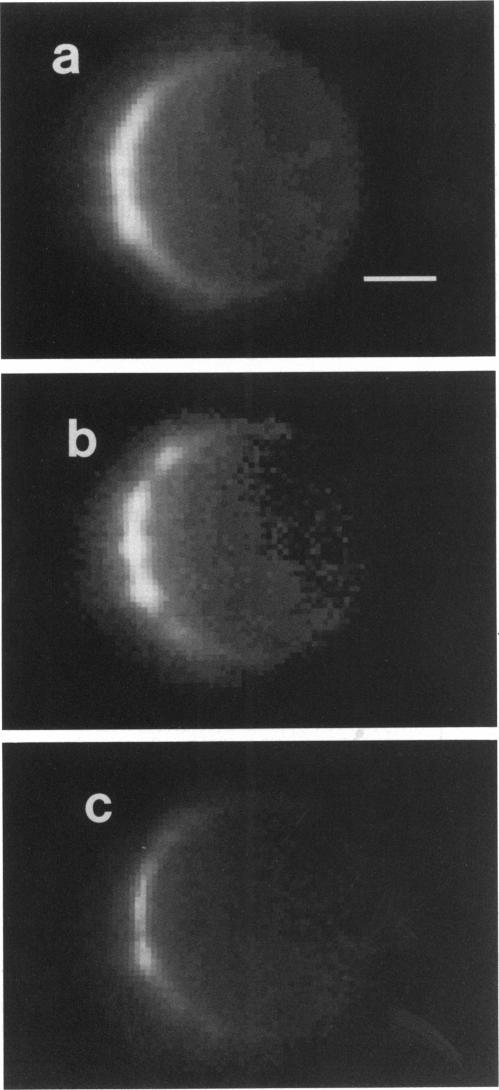
The ability of variations of membrane protein concentrations to modulate the lateral diffusion rate of an exemplary membrane protein has been studied in healthy and osmotically shocked cultured cells of the rat basophilic leukemia cell line, 2H3 subclone. Cell surface protein was redistributed by the method of in situ electrophoresis; exposure to electric fields of 1.25-5 V/cm results in cathodal migration of the majority of the surface proteins on this cell type (Ryan, T. A., J. Myers, D. Holowka, B. Baird, and W. W. Webb. Science [Wash. DC]. 239:61-64). Even in these small fields, the steady-state distribution becomes "crowded" with more than an 80% protein occupancy of accessible membrane area at the cathodal end of these spheroidal cells, and the anodal end becomes significantly depleted. We have employed fringe pattern fluorescence photobleaching with CCD imaging detection to measure lateral diffusion coefficients of the liganded IgE receptor on both crowded and uncrowded regions of individual rat basophilic leukemia cells. We find no significant difference in lateral diffusion rates in these regions. Cells swollen by hypoosmotic stress exhibit faster diffusion overall, with the uncrowded regions having a significantly greater increase in diffusion coefficient than the crowded regions. These results are consistent with the partial or total release of cytoskeletal constraints to membrane protein diffusion induced by osmotic stress.
在大鼠嗜碱性白血病细胞系2H3亚克隆的健康细胞和经渗透压休克处理的培养细胞中,研究了膜蛋白浓度变化调节一种示例性膜蛋白横向扩散速率的能力。通过原位电泳方法使细胞表面蛋白重新分布;暴露于1.25 - 5 V/cm的电场会导致这种细胞类型的大多数表面蛋白向阴极迁移(Ryan, T. A., J. Myers, D. Holowka, B. Baird, and W. W. Webb. Science [Wash. DC]. 239:61 - 64)。即使在这些小电场中,稳态分布也会变得“拥挤”,在这些球形细胞的阴极端,可及膜面积的蛋白占有率超过80%,而阳极端则明显耗尽。我们采用带电荷耦合器件(CCD)成像检测的条纹图案荧光光漂白技术来测量单个大鼠嗜碱性白血病细胞拥挤和不拥挤区域上结合的IgE受体的横向扩散系数。我们发现在这些区域的横向扩散速率没有显著差异。经低渗应激肿胀的细胞总体上表现出更快的扩散,不拥挤区域的扩散系数增加幅度明显大于拥挤区域。这些结果与渗透压应激诱导的细胞骨架对膜蛋白扩散的部分或完全约束释放相一致。