Dubois L, Landuyt W, Haustermans K, Dupont P, Bormans G, Vermaelen P, Flamen P, Verbeken E, Mortelmans L
Department of Nuclear Medicine, University Hospital Gasthuisberg and KU Leuven, Herestraat 49, 3000 Leuven, Belgium.
Br J Cancer. 2004 Nov 29;91(11):1947-54. doi: 10.1038/sj.bjc.6602219.
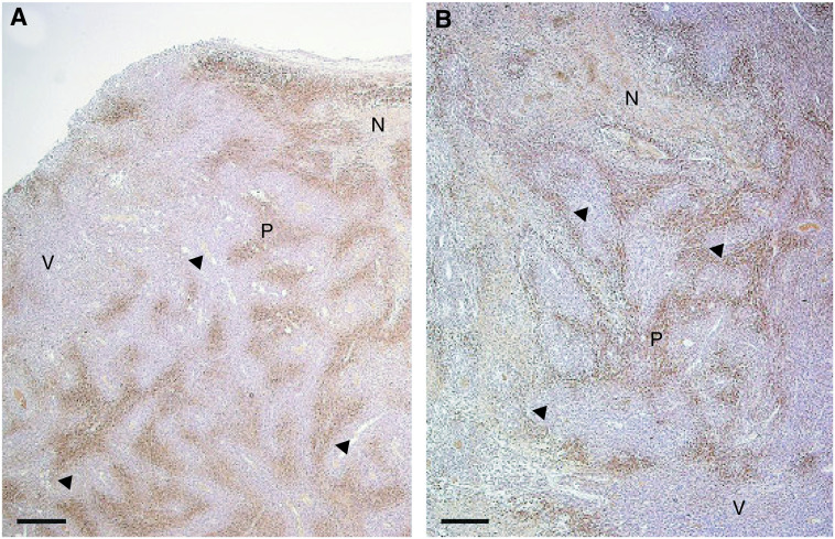
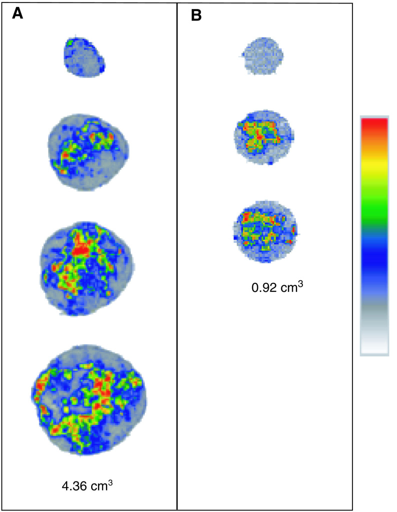
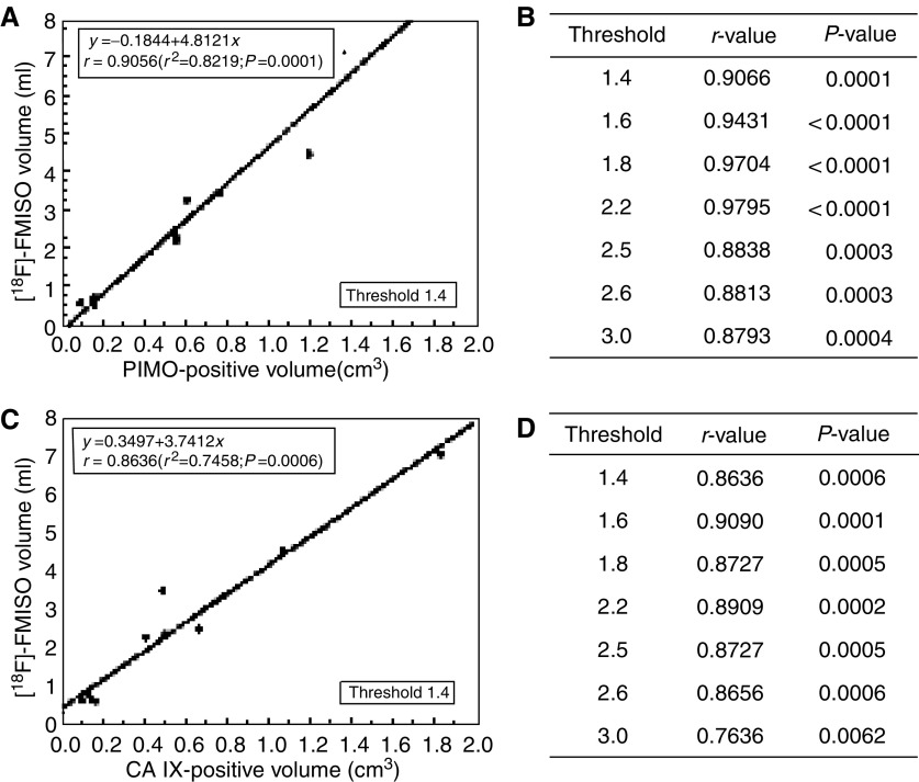
This study aimed to evaluate tumour hypoxia by comparing [(18)F]Fluoromisonidazole uptake measured using positron emission tomography ([(18)F]FMISO-PET) with immunohistochemical (IHC) staining techniques. Syngeneic rhabdomyosarcoma (R1) tumour pieces were transplanted subcutaneously in the flanks of WAG/Rij rats. Tumours were analysed at volumes between 0.9 and 7.3 cm(3). Hypoxic volumes were defined using a 3D region of interest on 2 h postinjection [(18)F]FMISO-PET images, applying different thresholds (1.2-3.0). Monoclonal antibodies to pimonidazole (PIMO) and carbonic anhydrase IX (CA IX), exogenous and endogenous markers of hypoxia, respectively, were used for IHC staining. Marker-positive fractions were microscopically measured for each tumour, and hypoxic volumes were calculated. A heterogeneous distribution of hypoxia was observed both with histology and [(18)F]FMISO autoradiography. A statistically significant correlation (P<0.05) was obtained between the hypoxic volumes defined with [(18)F]FMISO-PET and the volumes derived from the PIMO-stained tumour sections (r=0.9066; P=0.0001), regardless of the selected threshold between 1.4 and 2.2. A similar observation was made with the CA IX staining (r=0.8636; P=0.0006). The relationship found between [(18)F]FMISO-PET and PIMO- and additionally CA IX-derived hypoxic volumes in rat rhabdomyosarcomas indicates the value of the noninvasive imaging method to measure hypoxia in whole tumours.
本研究旨在通过比较使用正电子发射断层扫描([(18)F]FMISO-PET)测量的[(18)F]氟米索硝唑摄取与免疫组织化学(IHC)染色技术来评估肿瘤缺氧情况。将同基因横纹肌肉瘤(R1)肿瘤块皮下移植到WAG/Rij大鼠的侧腹。在肿瘤体积为0.9至7.3 cm(3)之间时对肿瘤进行分析。使用注射[(18)F]FMISO后2小时的PET图像上的三维感兴趣区域,应用不同阈值(1.2 - 3.0)来定义缺氧体积。分别针对缺氧的外源性和内源性标志物匹莫硝唑(PIMO)和碳酸酐酶IX(CA IX)的单克隆抗体用于IHC染色。在显微镜下测量每个肿瘤的标志物阳性分数,并计算缺氧体积。通过组织学和[(18)F]FMISO放射自显影均观察到缺氧的异质性分布。无论在1.4至2.2之间选择何种阈值,[(18)F]FMISO-PET定义的缺氧体积与PIMO染色肿瘤切片得出的体积之间均获得了统计学显著相关性(P<0.05)(r = 0.9066;P = 0.0001)。CA IX染色也有类似观察结果(r = 0.8636;P = 0.0006)。在大鼠横纹肌肉瘤中发现的[(18)F]FMISO-PET与PIMO以及另外CA IX衍生的缺氧体积之间的关系表明了这种非侵入性成像方法在测量整个肿瘤缺氧情况方面的价值。