• 文献检索
  • 文档翻译
  • 深度研究
  • 学术资讯
  • Suppr Zotero 插件Zotero 插件
  • 邀请有礼
  • 套餐&价格
  • 历史记录
应用&插件
Suppr Zotero 插件Zotero 插件浏览器插件Mac 客户端Windows 客户端微信小程序
定价
高级版会员购买积分包购买API积分包
服务
文献检索文档翻译深度研究API 文档MCP 服务
关于我们
关于 Suppr公司介绍联系我们用户协议隐私条款
关注我们

Suppr 超能文献

核心技术专利:CN118964589B侵权必究
粤ICP备2023148730 号-1Suppr @ 2026

文献检索

告别复杂PubMed语法,用中文像聊天一样搜索,搜遍4000万医学文献。AI智能推荐,让科研检索更轻松。

立即免费搜索

文件翻译

保留排版,准确专业,支持PDF/Word/PPT等文件格式,支持 12+语言互译。

免费翻译文档

深度研究

AI帮你快速写综述,25分钟生成高质量综述,智能提取关键信息,辅助科研写作。

立即免费体验

用于计算机辅助诊断中多图像分析的计算机化乳头识别

Computerized nipple identification for multiple image analysis in computer-aided diagnosis.

作者信息

Zhou Chuan, Chan Heang-Ping, Paramagul Chintana, Roubidoux Marilyn A, Sahiner Berkman, Hadjiiski Labomir M, Petrick Nicholas

机构信息

Department of Radiology, University of Michigan, Ann Arbor, Michigan 48109, USA.

出版信息

Med Phys. 2004 Oct;31(10):2871-82. doi: 10.1118/1.1800713.

DOI:10.1118/1.1800713
PMID:15543797
原文链接:https://pmc.ncbi.nlm.nih.gov/articles/PMC2898150/
Abstract

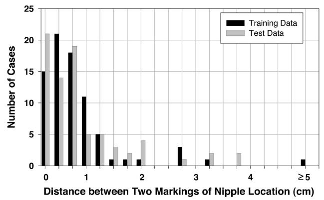
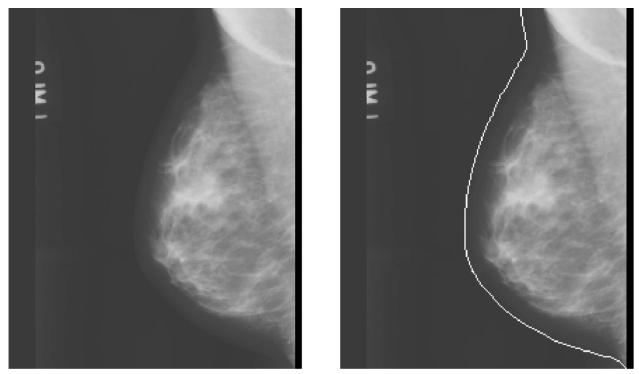
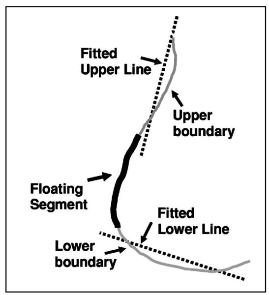
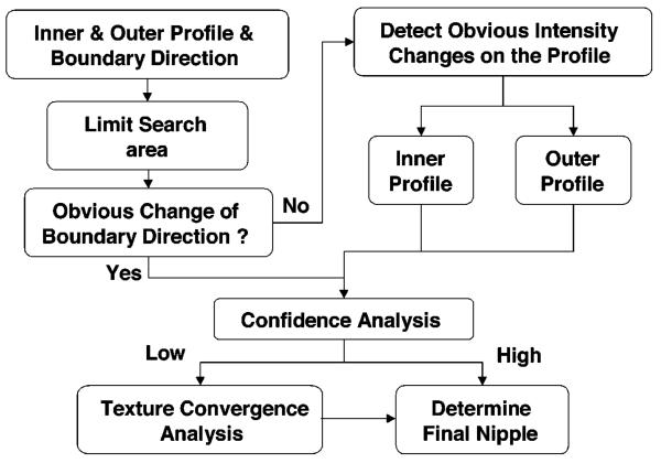
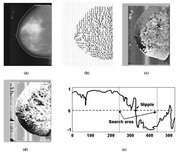
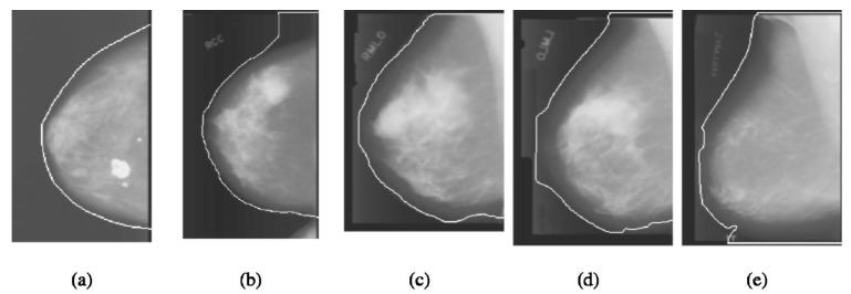
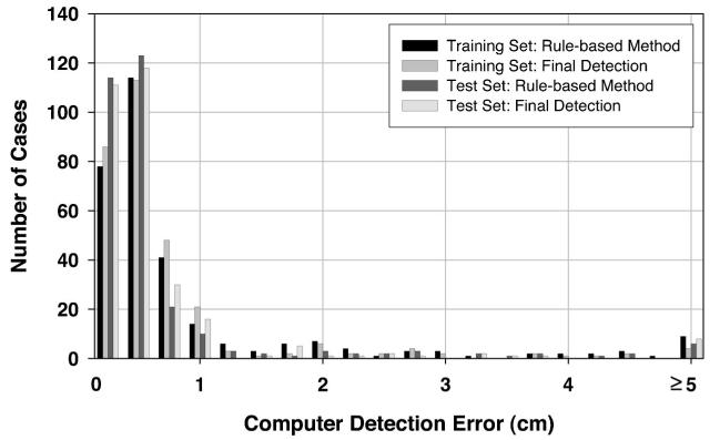
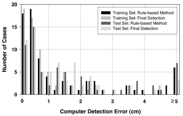
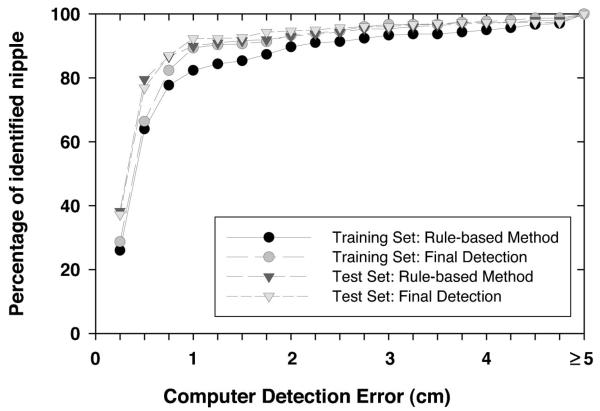
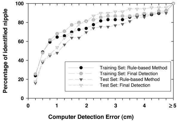
Correlation of information from multiple-view mammograms (e.g., MLO and CC views, bilateral views, or current and prior mammograms) can improve the performance of breast cancer diagnosis by radiologists or by computer. The nipple is a reliable and stable landmark on mammograms for the registration of multiple mammograms. However, accurate identification of nipple location on mammograms is challenging because of the variations in image quality and in the nipple projections, resulting in some nipples being nearly invisible on the mammograms. In this study, we developed a computerized method to automatically identify the nipple location on digitized mammograms. First, the breast boundary was obtained using a gradient-based boundary tracking algorithm, and then the gray level profiles along the inside and outside of the boundary were identified. A geometric convergence analysis was used to limit the nipple search to a region of the breast boundary. A two-stage nipple detection method was developed to identify the nipple location using the gray level information around the nipple, the geometric characteristics of nipple shapes, and the texture features of glandular tissue or ducts which converge toward the nipple. At the first stage, a rule-based method was designed to identify the nipple location by detecting significant changes of intensity along the gray level profiles inside and outside the breast boundary and the changes in the boundary direction. At the second stage, a texture orientation-field analysis was developed to estimate the nipple location based on the convergence of the texture pattern of glandular tissue or ducts towards the nipple. The nipple location was finally determined from the detected nipple candidates by a rule-based confidence analysis. In this study, 377 and 367 randomly selected digitized mammograms were used for training and testing the nipple detection algorithm, respectively. Two experienced radiologists identified the nipple locations which were used as the gold standard. In the training data set, 301 nipples were positively identified and were referred to as visible nipples. Seventy six nipples could not be positively identified and were referred to as invisible nipples. The radiologists provided their estimation of the nipple locations in the latter group for comparison with the computer estimates. The computerized method could detect 89.37% (269/301) of the visible nipples and 69.74% (53/76) of the invisible nipples within 1 cm of the gold standard. In the test data set, 298 and 69 of the nipples were classified as visible and invisible, respectively. 92.28% (275/298) of the visible nipples and 53.62% (37/69) of the invisible nipples were identified within 1 cm of the gold standard. The results demonstrate that the nipple locations on digitized mammograms can be accurately detected if they are visible and can be reasonably estimated if they are invisible. Automated nipple detection will be an important step towards multiple image analysis for CAD.

摘要

来自多个视角乳腺钼靶图像(例如,内外斜位和头尾位视图、双侧视图,或当前与之前的乳腺钼靶图像)的信息关联,可提高放射科医生或计算机进行乳腺癌诊断的性能。乳头是乳腺钼靶图像上用于配准多个乳腺钼靶图像的可靠且稳定的标志。然而,由于图像质量和乳头投影的变化,准确识别乳腺钼靶图像上的乳头位置具有挑战性,导致有些乳头在乳腺钼靶图像上几乎不可见。在本研究中,我们开发了一种计算机化方法来自动识别数字化乳腺钼靶图像上的乳头位置。首先,使用基于梯度的边界跟踪算法获取乳房边界,然后识别边界内外的灰度轮廓。采用几何收敛分析将乳头搜索限制在乳房边界的一个区域内。开发了一种两阶段乳头检测方法,利用乳头周围的灰度信息、乳头形状的几何特征以及向乳头汇聚的腺体组织或导管的纹理特征来识别乳头位置。在第一阶段,设计了一种基于规则的方法,通过检测乳房边界内外灰度轮廓上强度的显著变化以及边界方向的变化来识别乳头位置。在第二阶段,开发了一种纹理方向场分析方法,基于腺体组织或导管的纹理模式向乳头的汇聚来估计乳头位置。最后通过基于规则的置信度分析从检测到的乳头候选位置中确定乳头位置。在本研究中,分别使用377张和367张随机选择的数字化乳腺钼靶图像来训练和测试乳头检测算法。两名经验丰富的放射科医生确定乳头位置,将其用作金标准。在训练数据集中,301个乳头被明确识别,称为可见乳头。76个乳头未能被明确识别,称为不可见乳头。放射科医生对后一组乳头位置进行了估计,以便与计算机估计值进行比较。该计算机化方法能够在金标准的1厘米范围内检测出89.37%(269/301)的可见乳头和69.74%(53/76)的不可见乳头。在测试数据集中,分别有298个和69个乳头被分类为可见和不可见。在金标准的1厘米范围内识别出了92.28%(275/298)的可见乳头和53.62%(37/69)的不可见乳头。结果表明,数字化乳腺钼靶图像上的乳头位置如果可见则可以准确检测,如果不可见则可以合理估计。自动乳头检测将是朝着计算机辅助检测(CAD)的多图像分析迈出的重要一步。

https://cdn.ncbi.nlm.nih.gov/pmc/blobs/ab74/2898150/22e1e6d79d72/nihms-199849-f0011.jpg
https://cdn.ncbi.nlm.nih.gov/pmc/blobs/ab74/2898150/cab0ed6357a1/nihms-199849-f0001.jpg
https://cdn.ncbi.nlm.nih.gov/pmc/blobs/ab74/2898150/ab66fba61174/nihms-199849-f0002.jpg
https://cdn.ncbi.nlm.nih.gov/pmc/blobs/ab74/2898150/da420e589491/nihms-199849-f0003.jpg
https://cdn.ncbi.nlm.nih.gov/pmc/blobs/ab74/2898150/2cc222f614d0/nihms-199849-f0004.jpg
https://cdn.ncbi.nlm.nih.gov/pmc/blobs/ab74/2898150/97cb719f0da7/nihms-199849-f0005.jpg
https://cdn.ncbi.nlm.nih.gov/pmc/blobs/ab74/2898150/b9b9ec30eb38/nihms-199849-f0006.jpg
https://cdn.ncbi.nlm.nih.gov/pmc/blobs/ab74/2898150/80f43dc8dc9d/nihms-199849-f0007.jpg
https://cdn.ncbi.nlm.nih.gov/pmc/blobs/ab74/2898150/f3422c4d83a5/nihms-199849-f0008.jpg
https://cdn.ncbi.nlm.nih.gov/pmc/blobs/ab74/2898150/bc2414569e4b/nihms-199849-f0009.jpg
https://cdn.ncbi.nlm.nih.gov/pmc/blobs/ab74/2898150/91137cb1e84f/nihms-199849-f0010.jpg
https://cdn.ncbi.nlm.nih.gov/pmc/blobs/ab74/2898150/22e1e6d79d72/nihms-199849-f0011.jpg

相似文献

1
Computerized nipple identification for multiple image analysis in computer-aided diagnosis.用于计算机辅助诊断中多图像分析的计算机化乳头识别
Med Phys. 2004 Oct;31(10):2871-82. doi: 10.1118/1.1800713.
2
Fully automated nipple detection in digital breast tomosynthesis.数字乳腺断层合成中的全自动乳头检测
Comput Methods Programs Biomed. 2017 May;143:113-120. doi: 10.1016/j.cmpb.2017.03.004. Epub 2017 Mar 6.
3
A heuristic approach to automated nipple detection in digital mammograms.一种数字乳腺 X 线摄影中自动乳头检测的启发式方法。
J Digit Imaging. 2013 Oct;26(5):932-40. doi: 10.1007/s10278-013-9575-x.
4
A method to test the reproducibility and to improve performance of computer-aided detection schemes for digitized mammograms.一种用于测试数字化乳腺X线摄影计算机辅助检测方案的可重复性并提高其性能的方法。
Med Phys. 2004 Nov;31(11):2964-72. doi: 10.1118/1.1806291.
5
Dynamic multiple thresholding breast boundary detection algorithm for mammograms.动态多阈值乳腺边界检测算法在乳腺 X 线摄影中的应用。
Med Phys. 2010 Jan;37(1):391-401. doi: 10.1118/1.3273062.
6
An ellipse-fitting based method for efficient registration of breast masses on two mammographic views.一种基于椭圆拟合的方法,用于在两个乳腺钼靶视图上对乳腺肿块进行高效配准。
Med Phys. 2008 Feb;35(2):487-94. doi: 10.1118/1.2828188.
7
Automatic detection of the nipple in screen-film and full-field digital mammograms using a novel Hessian-based method.基于 Hessian 方法的屏片式和全数字化乳腺钼靶图像中自动检测乳头
J Digit Imaging. 2013 Oct;26(5):948-57. doi: 10.1007/s10278-013-9587-6.
8
Computer aided detection of clusters of microcalcifications on full field digital mammograms.全视野数字化乳腺钼靶片上微钙化簇的计算机辅助检测
Med Phys. 2006 Aug;33(8):2975-88. doi: 10.1118/1.2211710.
9
Automatic detection of breast border and nipple in digital mammograms.数字乳腺钼靶片中乳腺边界和乳头的自动检测。
Comput Methods Programs Biomed. 1996 May;49(3):253-62. doi: 10.1016/0169-2607(96)01724-5.
10
Dual system approach to computer-aided detection of breast masses on mammograms.乳腺钼靶片上乳腺肿块计算机辅助检测的双系统方法。
Med Phys. 2006 Nov;33(11):4157-68. doi: 10.1118/1.2357838.

引用本文的文献

1
Towards Automated Semantic Segmentation in Mammography Images for Enhanced Clinical Applications.迈向乳腺钼靶图像的自动语义分割以增强临床应用。
J Imaging Inform Med. 2024 Dec 11. doi: 10.1007/s10278-024-01364-8.
2
Mass detection in digital breast tomosynthesis: Deep convolutional neural network with transfer learning from mammography.数字乳腺断层合成中的肿块检测:基于乳腺X线摄影迁移学习的深度卷积神经网络
Med Phys. 2016 Dec;43(12):6654. doi: 10.1118/1.4967345.
3
Automatic detection of the nipple in screen-film and full-field digital mammograms using a novel Hessian-based method.

本文引用的文献

1
Filterbank-based fingerprint matching.基于滤波器组的指纹匹配。
IEEE Trans Image Process. 2000;9(5):846-59. doi: 10.1109/83.841531.
2
Improvement of computerized mass detection on mammograms: fusion of two-view information.乳腺钼靶X线摄影计算机辅助肿块检测的改进:双视图信息融合
Med Phys. 2002 Feb;29(2):238-47. doi: 10.1118/1.1446098.
3
Automated registration of breast lesions in temporal pairs of mammograms for interval change analysis--local affine transformation for improved localization.用于间隔期变化分析的乳腺钼靶影像时间序列中乳腺病变的自动配准——采用局部仿射变换以改进定位
基于 Hessian 方法的屏片式和全数字化乳腺钼靶图像中自动检测乳头
J Digit Imaging. 2013 Oct;26(5):948-57. doi: 10.1007/s10278-013-9587-6.
4
A heuristic approach to automated nipple detection in digital mammograms.一种数字乳腺 X 线摄影中自动乳头检测的启发式方法。
J Digit Imaging. 2013 Oct;26(5):932-40. doi: 10.1007/s10278-013-9575-x.
5
Computer-aided detection of breast masses: four-view strategy for screening mammography.计算机辅助检测乳房肿块:筛查性乳房 X 光摄影的四视图策略。
Med Phys. 2011 Apr;38(4):1867-76. doi: 10.1118/1.3560462.
6
Association of computerized mammographic parenchymal pattern measure with breast cancer risk: a pilot case-control study.计算机化乳腺组织密度测量与乳腺癌风险的关联:一项初步的病例对照研究。
Radiology. 2011 Jul;260(1):42-9. doi: 10.1148/radiol.11101266. Epub 2011 Mar 15.
7
Dynamic multiple thresholding breast boundary detection algorithm for mammograms.动态多阈值乳腺边界检测算法在乳腺 X 线摄影中的应用。
Med Phys. 2010 Jan;37(1):391-401. doi: 10.1118/1.3273062.
8
Computer-aided detection of breast masses on mammograms: dual system approach with two-view analysis.计算机辅助检测乳腺 X 线摄影中的肿块:双系统两视图分析方法。
Med Phys. 2009 Oct;36(10):4451-60. doi: 10.1118/1.3220669.
9
Application of boundary detection information in breast tomosynthesis reconstruction.边界检测信息在乳腺断层合成重建中的应用。
Med Phys. 2007 Sep;34(9):3603-13. doi: 10.1118/1.2761968.
10
Joint two-view information for computerized detection of microcalcifications on mammograms.联合双视图信息用于乳腺钼靶微钙化的计算机检测。
Med Phys. 2006 Jul;33(7):2574-85. doi: 10.1118/1.2208919.
Med Phys. 2001 Jun;28(6):1070-9. doi: 10.1118/1.1376134.
4
Computerized image analysis: estimation of breast density on mammograms.计算机图像分析:乳腺钼靶片上乳腺密度的估计
Med Phys. 2001 Jun;28(6):1056-69. doi: 10.1118/1.1376640.
5
Classification of compressed breast shapes for the design of equalization filters in x-ray mammography.用于乳腺X线摄影中均衡滤波器设计的压缩乳房形状分类
Med Phys. 1998 Jun;25(6):937-48. doi: 10.1118/1.598272.
6
Cancer statistics, 1998.1998年癌症统计数据。
CA Cancer J Clin. 1998 Jan-Feb;48(1):6-29. doi: 10.3322/canjclin.48.1.6.
7
A simple method for automatically locating the nipple on mammograms.一种在乳腺钼靶片上自动定位乳头的简单方法。
IEEE Trans Med Imaging. 1997 Oct;16(5):483-94. doi: 10.1109/42.640738.
8
Automatic detection of breast border and nipple in digital mammograms.数字乳腺钼靶片中乳腺边界和乳头的自动检测。
Comput Methods Programs Biomed. 1996 May;49(3):253-62. doi: 10.1016/0169-2607(96)01724-5.
9
Computerized detection of masses in digital mammograms: automated alignment of breast images and its effect on bilateral-subtraction technique.数字化乳腺钼靶片中肿块的计算机检测:乳腺图像的自动对齐及其对双侧相减技术的影响。
Med Phys. 1994 Mar;21(3):445-52. doi: 10.1118/1.597307.
10
Survival advantage differences by age. Evaluation of the extended follow-up of the Breast Cancer Detection Demonstration Project.按年龄划分的生存优势差异。乳腺癌检测示范项目延长随访的评估。
Cancer. 1994 Jul 1;74(1 Suppl):301-10. doi: 10.1002/cncr.2820741315.