Moon Jae-Youn, Chung Namsik, Choi Byoung Wook, Choe Kyu Ok, Seo Hye Sun, Ko Young-Guk, Kang Seok-Min, Ha Jong-Won, Rim Se-Joong, Jang Yangsoo, Shim Won-Heum, Cho Seung-Yun
Cardiology Division, Yonsei Cardiovascular Hospital, Yonsei University College of Medicine, 134 Shinchon-dong, Seodaemun-gu, Seoul 120-752, Korea.
Yonsei Med J. 2005 Feb 28;46(1):86-94. doi: 10.3349/ymj.2005.46.1.86.
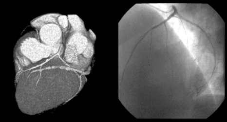
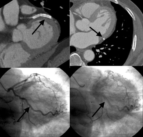
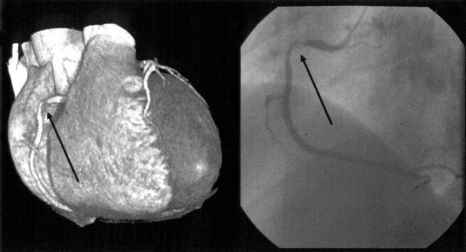
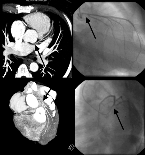
Contrast-enhanced multi-detector row spiral computed tomography (MDCT) was introduced as a promising noninvasive method for vascular imaging. This study examined the accuracy of this technique for detecting significant coronary artery stenoses. Both MDCT(Sensation 16, Siemens, Germany, 12x0.75 mm collimation and 0.42 sec rotation speed, 120 kV, 500 effective mA, and 2.7 mm/rotation table-feed) and invasive coronary angiography (CAG) were performed on 61 patients (mean age 59.2+/-10, 44 men) who were suspected of having coronary artery disease. All patients were treated with atenolol (25-50 mg) prior to imaging and the heart rate was maintained below 65 beats per minutes during image acquisition. The images were reconstructed in the diastole around TI-400 ms with a 0.5 mm increment and a 1.0 mm thickness. All coronary arteries with a diameter of 2.0 mm or more were assessed for the presence of a stenosis (>50% luminal narrowing). Two independent radiologists who were unaware of the results of the invasive CAG evaluated the MDCT data, and the results were compared with those from the invasive CAG (interval 1-27, mean 11 days). An evaluation of the CT coronary angiogram (CTCA) was possible in 58 of the 61 patients (95%). Image acquisition of the major coronary arteries including the left main trunk was available in 229 out of 244 arteries. Invasive CAG showed that 35 out of 58 patients had significant coronary artery stenoses by. patient analysis of those who could be evaluated showed that CT coronary angiography correctly classified 30 out of 35 patients as having at least 1 coronary stenosis (sensitivity 85.7%, specificity 91.3%, positive predictive value 93.8%, negative predictive value 80.8%). By analyzing each coronary artery, CAG found 62 stenotic coronary arteries in the 229 coronary arteries that could be evaluated. MDCT correctly detected 50 out of 62 stenotic coronary arteries and an absence of stenosis was correctly identified in 156 out of 167 normal coronary arteries (sensitivity 80.6%, specificity 93.4%, positive predictive value 81.9%, negative predictive value 92.8%). The non-invasive technique of MDCT for examining the coronary artery appears to be a useful method for detecting coronary artery stenoses with a high accuracy particularly with the proximal portion and large arteries.
对比增强型多排螺旋计算机断层扫描(MDCT)作为一种有前景的血管成像无创方法被引入。本研究检测了该技术检测显著冠状动脉狭窄的准确性。对61例疑似患有冠状动脉疾病的患者(平均年龄59.2±10岁,44例男性)同时进行了MDCT(德国西门子Sensation 16,准直12×0.75mm,转速0.42秒,120kV,有效毫安500,床进速度2.7mm/旋转)和有创冠状动脉造影(CAG)检查。所有患者在成像前均接受阿替洛尔(25 - 50mg)治疗,且在图像采集期间心率维持在每分钟65次以下。图像在舒张期TI - 400ms左右重建,增量为0.5mm,层厚为1.0mm。对所有直径2.0mm及以上的冠状动脉评估是否存在狭窄(管腔狭窄>50%)。两名不知有创CAG结果的独立放射科医生评估MDCT数据,并将结果与有创CAG的结果进行比较(间隔1 - 27天,平均11天)。61例患者中有58例(95%)可行CT冠状动脉造影(CTCA)评估。244支动脉中有229支获得了包括左主干在内的主要冠状动脉的图像采集。有创CAG显示,58例患者中有35例存在显著冠状动脉狭窄。对可评估患者的分析显示,CT冠状动脉造影将35例患者中的30例正确分类为至少有1处冠状动脉狭窄(敏感性85.7%,特异性91.3%,阳性预测值93.8%,阴性预测值80.8%)。通过分析每支冠状动脉,CAG在229支可评估的冠状动脉中发现62支狭窄冠状动脉。MDCT正确检测出62支狭窄冠状动脉中的50支,在167支正常冠状动脉中的156支正确识别出无狭窄(敏感性80.6%,特异性93.4%,阳性预测值81.9%,阴性预测值92.8%)。MDCT这种用于检查冠状动脉的无创技术似乎是一种检测冠状动脉狭窄的有用方法,尤其对于近端部分和大动脉具有较高的准确性。