• 文献检索
  • 文档翻译
  • 深度研究
  • 学术资讯
  • Suppr Zotero 插件Zotero 插件
  • 邀请有礼
  • 套餐&价格
  • 历史记录
应用&插件
Suppr Zotero 插件Zotero 插件浏览器插件Mac 客户端Windows 客户端微信小程序
定价
高级版会员购买积分包购买API积分包
服务
文献检索文档翻译深度研究API 文档MCP 服务
关于我们
关于 Suppr公司介绍联系我们用户协议隐私条款
关注我们

Suppr 超能文献

核心技术专利:CN118964589B侵权必究
粤ICP备2023148730 号-1Suppr @ 2026

文献检索

告别复杂PubMed语法,用中文像聊天一样搜索,搜遍4000万医学文献。AI智能推荐,让科研检索更轻松。

立即免费搜索

文件翻译

保留排版,准确专业,支持PDF/Word/PPT等文件格式,支持 12+语言互译。

免费翻译文档

深度研究

AI帮你快速写综述,25分钟生成高质量综述,智能提取关键信息,辅助科研写作。

立即免费体验

利用多光子显微镜在体内对肿瘤中的绿色荧光蛋白(GFP)、青色荧光蛋白(CFP)和胶原蛋白进行同步成像。

Simultaneous imaging of GFP, CFP and collagen in tumors in vivo using multiphoton microscopy.

作者信息

Sahai Erik, Wyckoff Jeffrey, Philippar Ulrike, Segall Jeffrey E, Gertler Frank, Condeelis John

机构信息

Tumour Cell Biology Laboratory, Cancer Research UK London Research Institute, London WC2A 3PX, UK.

出版信息

BMC Biotechnol. 2005 May 23;5:14. doi: 10.1186/1472-6750-5-14.

DOI:10.1186/1472-6750-5-14
PMID:15910685
原文链接:https://pmc.ncbi.nlm.nih.gov/articles/PMC1159039/
Abstract

BACKGROUND

The development of multiphoton laser scanning microscopy has greatly facilitated the imaging of living tissues. However, the use of genetically encoded fluorescent proteins to distinguish different cell types in living animals has not been described at single cell resolution using multiphoton microscopy.

RESULTS

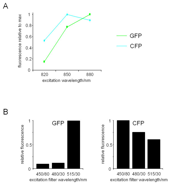
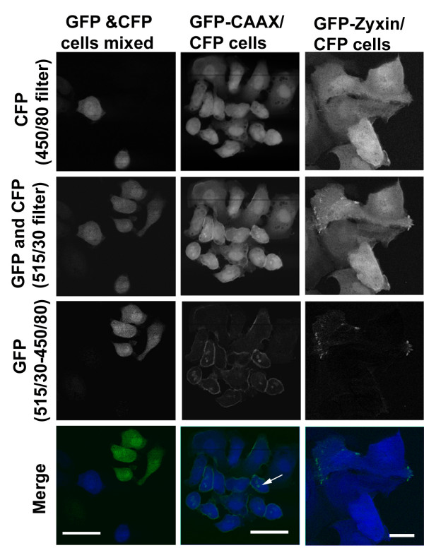
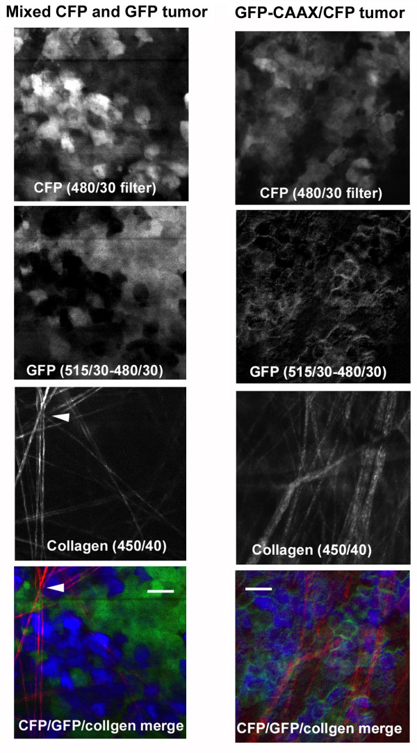
Here we describe a method for the simultaneous imaging, by multiphoton microscopy, of Green Fluorescent Protein, Cyan Fluorescent Protein and collagen in vivo in living tumors. This novel method enables: 1) the simultaneous visualization of overall cell shape and sub-cellular structures such as the plasma membrane or proteins of interest in cells inside living animals, 2) direct comparison of the behavior of single cells from different cell lines in the same microenvironment in vivo.

CONCLUSION

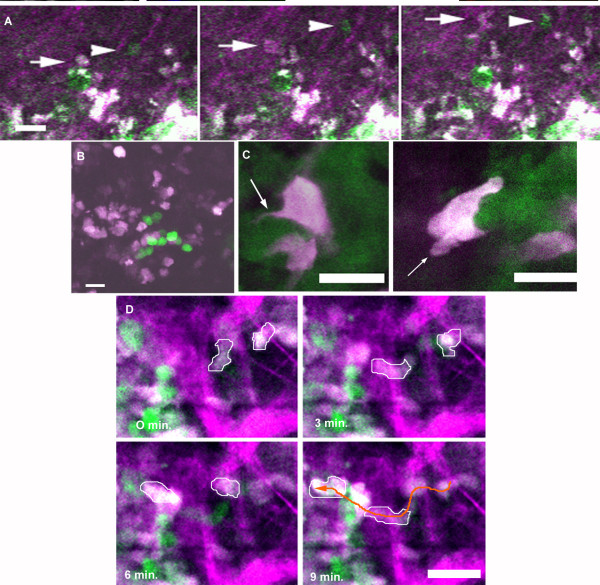
Using this multi-fluor, multiphoton technique, we demonstrate that motility and metastatic differences between carcinoma cells of differing metastatic potential can be imaged in the same animal simultaneously at sub-cellular resolution.

摘要

背景

多光子激光扫描显微镜的发展极大地促进了对活组织的成像。然而,尚未有研究利用多光子显微镜在单细胞分辨率下描述使用基因编码荧光蛋白来区分活体动物中不同细胞类型的情况。

结果

在此,我们描述了一种通过多光子显微镜在活体肿瘤体内同时成像绿色荧光蛋白、青色荧光蛋白和胶原蛋白的方法。这种新方法能够:1)在活体动物体内同时可视化整体细胞形态和亚细胞结构,如质膜或细胞内感兴趣的蛋白质;2)直接比较来自不同细胞系的单个细胞在体内同一微环境中的行为。

结论

使用这种多荧光、多光子技术,我们证明了具有不同转移潜能的癌细胞之间的运动性和转移差异能够在同一动物体内以亚细胞分辨率同时成像。

https://cdn.ncbi.nlm.nih.gov/pmc/blobs/e87e/1159039/47ff3a734e5b/1472-6750-5-14-4.jpg
https://cdn.ncbi.nlm.nih.gov/pmc/blobs/e87e/1159039/a0a0fb44cf24/1472-6750-5-14-1.jpg
https://cdn.ncbi.nlm.nih.gov/pmc/blobs/e87e/1159039/9ac327bf7f8e/1472-6750-5-14-2.jpg
https://cdn.ncbi.nlm.nih.gov/pmc/blobs/e87e/1159039/d6d8b187bc76/1472-6750-5-14-3.jpg
https://cdn.ncbi.nlm.nih.gov/pmc/blobs/e87e/1159039/47ff3a734e5b/1472-6750-5-14-4.jpg

相似文献

1
Simultaneous imaging of GFP, CFP and collagen in tumors in vivo using multiphoton microscopy.利用多光子显微镜在体内对肿瘤中的绿色荧光蛋白(GFP)、青色荧光蛋白(CFP)和胶原蛋白进行同步成像。
BMC Biotechnol. 2005 May 23;5:14. doi: 10.1186/1472-6750-5-14.
2
Intact corneal stroma visualization of GFP mouse revealed by multiphoton imaging.通过多光子成像揭示的绿色荧光蛋白小鼠完整角膜基质可视化。
Microsc Res Tech. 2006 Dec;69(12):973-5. doi: 10.1002/jemt.20373.
3
Probing the microenvironment of mammary tumors using multiphoton microscopy.使用多光子显微镜探究乳腺肿瘤的微环境。
J Mammary Gland Biol Neoplasia. 2006 Apr;11(2):151-63. doi: 10.1007/s10911-006-9021-5.
4
Multiphoton tomography visualizes collagen fibers in the tumor microenvironment that maintain cancer-cell anchorage and shape.多光子断层成像技术可可视化肿瘤微环境中的胶原纤维,这些纤维维持着癌细胞的附着和形状。
J Cell Biochem. 2013 Jan;114(1):99-102. doi: 10.1002/jcb.24305.
5
Multiphoton flow cytometry strategies and applications.多光子流式细胞术策略及其应用。
Cytometry A. 2011 Oct;79(10):775-88. doi: 10.1002/cyto.a.21110. Epub 2011 Jul 27.
6
Imaging tumor cell movement in vivo.体内肿瘤细胞运动成像。
Curr Protoc Cell Biol. 2007 Jun;Chapter 19:Unit 19.7. doi: 10.1002/0471143030.cb1907s35.
7
In vivo real-time imaging of chemotherapy response on the liver metastatic tumor microenvironment using multiphoton microscopy.利用多光子显微镜对肝转移瘤微环境中的化疗反应进行体内实时成像。
Oncol Rep. 2012 Nov;28(5):1822-30. doi: 10.3892/or.2012.1983. Epub 2012 Aug 23.
8
The cyan fluorescent protein nude mouse as a host for multicolor-coded imaging models of primary and metastatic tumor microenvironments.作为一种宿主,青色荧光蛋白裸鼠可用于原发性和转移性肿瘤微环境的多色编码成像模型。
Anticancer Res. 2012 Jan;32(1):31-8.
9
Visualization of intrathoracically disseminated solid tumors in mice with optical imaging by telomerase-specific amplification of a transferred green fluorescent protein gene.通过对转移的绿色荧光蛋白基因进行端粒酶特异性扩增,利用光学成像技术在小鼠体内可视化胸腔内播散的实体瘤。
Cancer Res. 2004 Sep 1;64(17):6259-65. doi: 10.1158/0008-5472.CAN-04-1335.
10
Cancer-cell killing by engineered Salmonella imaged by multiphoton tomography in live mice.工程化沙门氏菌在活体小鼠中通过多光子断层成像杀死癌细胞。
Anticancer Res. 2012 Oct;32(10):4331-7.

引用本文的文献

1
Pathological and molecular insights into intravenous leiomyomatosis: an integrative multi-omics study.静脉内平滑肌瘤病的病理学和分子见解:一项综合多组学研究。
J Transl Med. 2025 Feb 26;23(1):229. doi: 10.1186/s12967-024-05919-9.
2
The Multifaced Role of Collagen in Cancer Development and Progression.胶原蛋白在癌症发生发展中的多面作用
Int J Mol Sci. 2024 Dec 17;25(24):13523. doi: 10.3390/ijms252413523.
3
Uncovering Novel Protein Partners of Inducible Nitric Oxide Synthase in Human Testis.揭示人睾丸中诱导型一氧化氮合酶的新型蛋白质伴侣。

本文引用的文献

1
Single-photon counting multicolor multiphoton fluorescence microscope.单光子计数多色多光子荧光显微镜
J Fluoresc. 2005 Jan;15(1):41-51. doi: 10.1007/s10895-005-0212-z.
2
FRET multiphoton spectral imaging microscopy of 7-ketocholesterol and Nile Red in U937 monocytic cells loaded with 7-ketocholesterol.对加载了7-酮胆固醇的U937单核细胞中的7-酮胆固醇和尼罗红进行荧光共振能量转移多光子光谱成像显微镜观察。
Anal Quant Cytol Histol. 2004 Dec;26(6):304-13.
3
Imaging calpain protease activity by multiphoton FRET in living mice.在活体小鼠中通过多光子荧光共振能量转移成像检测钙蛋白酶活性
Biomolecules. 2024 Mar 24;14(4):388. doi: 10.3390/biom14040388.
4
The impact of tumor microenvironment: unraveling the role of physical cues in breast cancer progression.肿瘤微环境的影响:揭示物理线索在乳腺癌进展中的作用。
Cancer Metastasis Rev. 2024 Jun;43(2):823-844. doi: 10.1007/s10555-024-10166-x. Epub 2024 Jan 19.
5
Collagens in Cancer: Structural Regulators and Guardians of Cancer Progression.癌症中的胶原蛋白:结构调节剂和癌症进展的守护者。
Cancer Res. 2023 May 2;83(9):1386-1392. doi: 10.1158/0008-5472.CAN-22-2034.
6
Assessment of MRI to estimate metastatic dissemination risk and prometastatic effects of chemotherapy.评估磁共振成像以估计转移扩散风险及化疗的促转移作用。
NPJ Breast Cancer. 2022 Sep 2;8(1):101. doi: 10.1038/s41523-022-00463-5.
7
Real-time polarization microscopy of fibrillar collagen in histopathology.组织病理学中纤维状胶原的实时偏光显微镜观察。
Sci Rep. 2021 Sep 24;11(1):19063. doi: 10.1038/s41598-021-98600-w.
8
Intravital microscopy of dynamic single-cell behavior in mouse mammary tissue.活体显微镜观察小鼠乳腺组织中动态单细胞行为。
Nat Protoc. 2021 Apr;16(4):1907-1935. doi: 10.1038/s41596-020-00473-2. Epub 2021 Feb 24.
9
Mammographic density: intersection of advocacy, science, and clinical practice.乳腺钼靶密度:倡导、科学与临床实践的交叉点。
Curr Breast Cancer Rep. 2019 Sep;11(3):100-110. doi: 10.1007/s12609-019-00316-4. Epub 2019 Jul 24.
10
Intratumoral collagen signatures predict clinical outcomes in feline mammary carcinoma.肿瘤内胶原特征可预测猫乳腺肿瘤的临床结局。
PLoS One. 2020 Aug 10;15(8):e0236516. doi: 10.1371/journal.pone.0236516. eCollection 2020.
J Mol Biol. 2005 Feb 11;346(1):215-22. doi: 10.1016/j.jmb.2004.11.039. Epub 2004 Dec 16.
4
Identification and testing of a gene expression signature of invasive carcinoma cells within primary mammary tumors.原发性乳腺肿瘤中侵袭性癌细胞基因表达特征的鉴定与检测。
Cancer Res. 2004 Dec 1;64(23):8585-94. doi: 10.1158/0008-5472.CAN-04-1136.
5
Interpreting second-harmonic generation images of collagen I fibrils.解读I型胶原纤维的二次谐波产生图像。
Biophys J. 2005 Feb;88(2):1377-86. doi: 10.1529/biophysj.104.047308. Epub 2004 Nov 8.
6
Intravital imaging of cell movement in tumours.肿瘤中细胞运动的活体成像
Nat Rev Cancer. 2003 Dec;3(12):921-30. doi: 10.1038/nrc1231.
7
Determination of clonality of metastasis by cell-specific color-coded fluorescent-protein imaging.通过细胞特异性颜色编码荧光蛋白成像确定转移灶的克隆性
Cancer Res. 2003 Nov 15;63(22):7785-90.
8
Dual-color fluorescence imaging distinguishes tumor cells from induced host angiogenic vessels and stromal cells.双色荧光成像可区分肿瘤细胞与诱导生成的宿主血管生成血管及基质细胞。
Proc Natl Acad Sci U S A. 2003 Nov 25;100(24):14259-62. doi: 10.1073/pnas.2436101100. Epub 2003 Nov 12.
9
Nonlinear magic: multiphoton microscopy in the biosciences.非线性魔法:生物科学中的多光子显微镜术
Nat Biotechnol. 2003 Nov;21(11):1369-77. doi: 10.1038/nbt899.
10
Viable circulating metastatic cells produced in orthotopic but not ectopic prostate cancer models.原位而非异位前列腺癌模型中产生了有活力的循环转移细胞。
Cancer Res. 2003 Jul 15;63(14):4239-43.