• 文献检索
  • 文档翻译
  • 深度研究
  • 学术资讯
  • Suppr Zotero 插件Zotero 插件
  • 邀请有礼
  • 套餐&价格
  • 历史记录
应用&插件
Suppr Zotero 插件Zotero 插件浏览器插件Mac 客户端Windows 客户端微信小程序
定价
高级版会员购买积分包购买API积分包
服务
文献检索文档翻译深度研究API 文档MCP 服务
关于我们
关于 Suppr公司介绍联系我们用户协议隐私条款
关注我们

Suppr 超能文献

核心技术专利:CN118964589B侵权必究
粤ICP备2023148730 号-1Suppr @ 2026

文献检索

告别复杂PubMed语法,用中文像聊天一样搜索,搜遍4000万医学文献。AI智能推荐,让科研检索更轻松。

立即免费搜索

文件翻译

保留排版,准确专业,支持PDF/Word/PPT等文件格式,支持 12+语言互译。

免费翻译文档

深度研究

AI帮你快速写综述,25分钟生成高质量综述,智能提取关键信息,辅助科研写作。

立即免费体验

肺癌患者无大量胸腔积液的细微胸膜转移:CT术前检测

Subtle pleural metastasis without large effusion in lung cancer patients: preoperative detection on CT.

作者信息

Hwang Jung Hwa, Song Koun-Sik, Park Seung-Il, Lim Tae-Hwan, Kwon Kui Hyang, Goo Dong Erk

机构信息

Department of Radiology, Soonchunhyang University Hospital, Seoul, Korea.

出版信息

Korean J Radiol. 2005 Apr-Jun;6(2):94-101. doi: 10.3348/kjr.2005.6.2.94.

DOI:10.3348/kjr.2005.6.2.94
PMID:15968148
原文链接:https://pmc.ncbi.nlm.nih.gov/articles/PMC2686426/
Abstract

OBJECTIVE

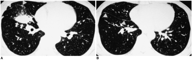
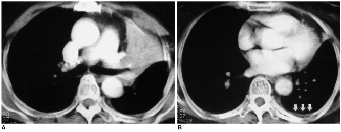
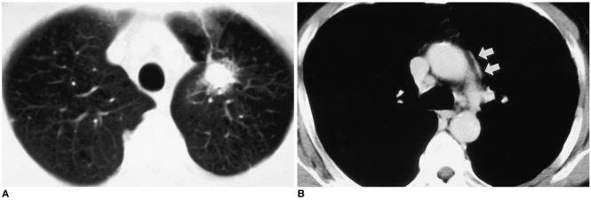
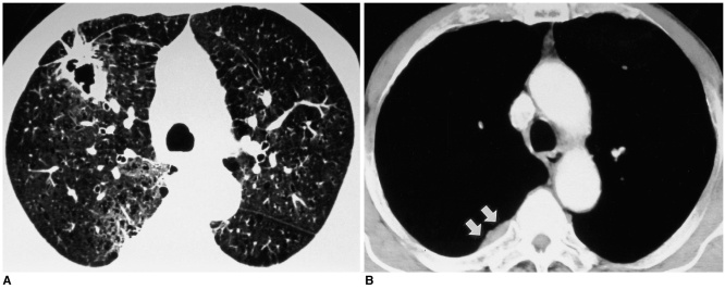
We wanted to describe the retrospective CT features of subtle pleural metastasis without large effusion that would suggest inoperable lung cancer.

MATERIALS AND METHODS

We enrolled 14 patients who had open thoracotomy attempted for lung cancer, but they were proven to be inoperable due to pleural metastasis. Our study also included 20 control patients who were proven as having no pleural metastasis. We retrospectively evaluated the nodularity and thickening of the pleura and the associated pleural effusion on the preoperative chest CT scans. We reviewed the histologic cancer types, the size, shape and location of the lung cancer and the associated mediastinal lymphadenopathy.

RESULTS

Subtle pleural nodularity or focal thickening was noted in seven patients (50%) having pleural metastasis and also in three patients (15%) of control group who were without pleural metastasis. More than one of the pleural changes such as subtle pleural nodularity, focal thickening or effusion was identified in eight (57%) patients having pleural metastasis and also in three patients (15%) of the control group, and these findings were significantly less frequent in the control group patients than for the patients with pleural metastasis (p = 0.02). The histologic types of primary lung cancer in patients with pleural metastasis revealed as adenocarcinoma in 10 patients (71%) and squamous cell carcinoma in four patients (29%). The location, size and shape of the primary lung cancer and the associated mediastinal lymphadenopathy showed no significant correlation with pleural metastasis.

CONCLUSION

If any subtle pleural nodularity or thickening is found on preoperative chest CT scans of patients with lung cancer, the possibility of pleural metastasis should be considered.

摘要

目的

我们旨在描述无大量胸腔积液的细微胸膜转移的回顾性CT特征,此类特征提示肺癌无法手术切除。

材料与方法

我们纳入了14例因肺癌尝试进行开胸手术但因胸膜转移而被证明无法手术的患者。我们的研究还包括20例被证明无胸膜转移的对照患者。我们回顾性评估了术前胸部CT扫描中胸膜的结节状和增厚情况以及相关的胸腔积液。我们复查了组织学癌症类型、肺癌的大小、形状和位置以及相关的纵隔淋巴结肿大情况。

结果

7例(50%)有胸膜转移的患者以及3例(15%)无胸膜转移的对照组患者出现了细微胸膜结节或局灶性增厚。8例(57%)有胸膜转移的患者以及3例(15%)对照组患者出现了不止一种胸膜改变,如细微胸膜结节、局灶性增厚或胸腔积液,并且这些发现在对照组患者中的出现频率显著低于胸膜转移患者(p = 0.02)。胸膜转移患者中原发性肺癌的组织学类型显示,10例(71%)为腺癌,4例(29%)为鳞状细胞癌。原发性肺癌的位置、大小和形状以及相关的纵隔淋巴结肿大与胸膜转移无显著相关性。

结论

如果在肺癌患者的术前胸部CT扫描中发现任何细微胸膜结节或增厚,应考虑胸膜转移的可能性。

https://cdn.ncbi.nlm.nih.gov/pmc/blobs/a6fe/2686426/0786801d5e77/kjr-6-94-g004.jpg
https://cdn.ncbi.nlm.nih.gov/pmc/blobs/a6fe/2686426/e658de9083a1/kjr-6-94-g001.jpg
https://cdn.ncbi.nlm.nih.gov/pmc/blobs/a6fe/2686426/a357cbd4b897/kjr-6-94-g002.jpg
https://cdn.ncbi.nlm.nih.gov/pmc/blobs/a6fe/2686426/eaaac57369f5/kjr-6-94-g003.jpg
https://cdn.ncbi.nlm.nih.gov/pmc/blobs/a6fe/2686426/0786801d5e77/kjr-6-94-g004.jpg

相似文献

1
Subtle pleural metastasis without large effusion in lung cancer patients: preoperative detection on CT.肺癌患者无大量胸腔积液的细微胸膜转移:CT术前检测
Korean J Radiol. 2005 Apr-Jun;6(2):94-101. doi: 10.3348/kjr.2005.6.2.94.
2
Pleural and chest wall invasion in bronchogenic carcinoma: CT evaluation.支气管肺癌的胸膜及胸壁侵犯:CT评估
Radiology. 1985 Oct;157(1):191-4. doi: 10.1148/radiology.157.1.4034965.
3
[Thoracocentesis for the assessment of lung cancer with pleural effusion].[胸腔穿刺术用于评估伴有胸腔积液的肺癌]
Arch Bronconeumol. 2002 Oct;38(10):479-84. doi: 10.1016/s0300-2896(02)75269-1.
4
[Diagnosis of pleural dissemination lung cancer by thin-section CT--a prospective study].[薄层CT诊断胸膜播散性肺癌的前瞻性研究]
Nihon Kyobu Shikkan Gakkai Zasshi. 1996 Nov;34(11):1194-201.
5
Carcinoma of the lung in HIV-positive patients: findings on chest radiographs and CT scans.
AJR Am J Roentgenol. 1995 Mar;164(3):593-7. doi: 10.2214/ajr.164.3.7863877.
6
Dry small pleural dissemination of adenocarcinoma of the lung preoperatively detected by PET/CT: a report of two cases.PET/CT术前检测到的肺腺癌干性小胸膜播散:2例报告
Acta Med Okayama. 2008 Feb;62(1):55-8. doi: 10.18926/AMO/30991.
7
[Pleural and parenchymal lung diseases from asbestos exposure. CT diagnosis].[石棉暴露所致胸膜和肺实质疾病。CT诊断]
Radiol Med. 2000 Nov;100(5):326-31.
8
CT diagnosis of pleural dissemination without pleural effusion in primary lung cancer.原发性肺癌无胸腔积液的胸膜播散的CT诊断
Radiat Med. 1996 May-Jun;14(3):117-9.
9
Diagnostic Value of Computed Tomography Imaging Features in Malignant Pleural Mesothelioma.计算机断层扫描成像特征对恶性胸膜间皮瘤的诊断价值。
Respiration. 2020;99(1):28-34. doi: 10.1159/000503239. Epub 2019 Oct 25.
10
Minimal Pleural Effusion in Small Cell Lung Cancer: Proportion, Mechanisms, and Prognostic Effect.小细胞肺癌中的少量胸腔积液:比例、机制和预后影响。
Radiology. 2016 Feb;278(2):593-600. doi: 10.1148/radiol.2015142388. Epub 2015 Aug 31.

引用本文的文献

1
Enhancing Intrapleural Hyperthermic Chemotherapy for Lung Cancer: Insights from 3D and PDX Models.增强肺癌的胸腔内热化疗:来自三维和人源肿瘤异种移植模型的见解
Cancers (Basel). 2024 Oct 11;16(20):3448. doi: 10.3390/cancers16203448.
2
[Pleural Metastasis of Lung Cancer Combined with Pleuroparenchymal Fibroelastosis: A Case Report].[肺癌胸膜转移合并胸膜实质纤维弹性组织增生症:1例报告]
J Korean Soc Radiol. 2024 May;85(3):631-636. doi: 10.3348/jksr.2023.0024. Epub 2024 Mar 8.
3
Added value of 40 keV virtual monoenergetic images for diagnosing malignant pleural effusion on chest CT.

本文引用的文献

1
Staging of non-small-cell lung cancer with integrated positron-emission tomography and computed tomography.正电子发射断层扫描与计算机断层扫描相结合用于非小细胞肺癌分期
N Engl J Med. 2003 Jun 19;348(25):2500-7. doi: 10.1056/NEJMoa022136.
2
Helical computed tomography diagnosis of pleural dissemination in lung cancer: comparison of thick-section and thin-section helical computed tomography.螺旋计算机断层扫描对肺癌胸膜播散的诊断:厚层与薄层螺旋计算机断层扫描的比较
J Thorac Imaging. 1998 Jul;13(3):211-8. doi: 10.1097/00005382-199807000-00009.
3
Pleural cytologies in lung cancer without pleural effusions.
40keV 虚拟单能量图像在胸部 CT 诊断恶性胸腔积液中的附加价值。
Jpn J Radiol. 2024 Aug;42(8):862-871. doi: 10.1007/s11604-024-01571-x. Epub 2024 May 3.
4
Predicting pleural invasion of invasive lung adenocarcinoma in the adjacent pleura by imaging histology.通过影像组织学预测侵袭性肺腺癌在邻近胸膜中的胸膜侵犯情况。
Oncol Lett. 2023 Aug 23;26(4):438. doi: 10.3892/ol.2023.14025. eCollection 2023 Oct.
5
Differentiating malignant and benign pleural effusion in patients with lung cancer: an F-FDG PET/CT retrospectively study.肺癌患者恶性与良性胸腔积液的鉴别:一项F-FDG PET/CT回顾性研究。
Front Oncol. 2023 Jun 30;13:1192870. doi: 10.3389/fonc.2023.1192870. eCollection 2023.
6
The Role of 18F-FDG PET/CT Integrated Imaging in Distinguishing Malignant from Benign Pleural Effusion.18F-FDG PET/CT融合成像在鉴别恶性与良性胸腔积液中的作用
PLoS One. 2016 Aug 25;11(8):e0161764. doi: 10.1371/journal.pone.0161764. eCollection 2016.
7
Association of epidermal growth factor receptor mutations with metastatic presentations in non-small cell lung cancer.表皮生长因子受体突变与非小细胞肺癌转移表现的相关性
ISRN Oncol. 2011;2011:756265. doi: 10.5402/2011/756265. Epub 2011 Apr 27.
8
Integrated imaging of non-small cell lung cancer recurrence: CT and PET-CT findings, possible pitfalls and risk of recurrence criteria.非小细胞肺癌复发的综合影像:CT 和 PET-CT 表现、可能的陷阱和复发标准的风险。
Eur Radiol. 2012 Mar;22(3):588-606. doi: 10.1007/s00330-011-2299-8. Epub 2011 Oct 4.
无胸腔积液的肺癌患者的胸膜细胞学检查
Ann Thorac Surg. 1997 Oct;64(4):941-4. doi: 10.1016/s0003-4975(97)00817-5.
4
Thoracoscopic evaluation of histologically/cytologically proven or suspected lung cancer: a VATS exploration.经组织学/细胞学证实或怀疑为肺癌的胸腔镜评估:一项电视辅助胸腔镜手术探查
Lung Cancer. 1997 Mar;16(2-3):183-90. doi: 10.1016/s0169-5002(96)00628-9.
5
CT diagnosis of pleural dissemination without pleural effusion in primary lung cancer.原发性肺癌无胸腔积液的胸膜播散的CT诊断
Radiat Med. 1996 May-Jun;14(3):117-9.
6
Surgical experience in treating T4 lung cancer: its resectability, morbidity, mortality and prognosis.T4期肺癌的外科治疗经验:可切除性、发病率、死亡率及预后
Eur J Surg Oncol. 1996 Apr;22(2):171-6. doi: 10.1016/s0748-7983(96)90775-1.
7
Comparison of pleuropneumonectomy and limited surgery for lung cancer with pleural dissemination.肺癌伴胸膜播散的胸膜肺切除术与有限手术的比较。
J Surg Oncol. 1996 Jan;61(1):1-6. doi: 10.1002/(SICI)1096-9098(199601)61:1<1::AID-JSO1>3.0.CO;2-H.
8
Thoracoscopic surgery for diseases of the lung and pleura. Effectiveness, changing indications, and limitations.用于治疗肺部和胸膜疾病的胸腔镜手术。有效性、适应证的变化及局限性。
Ann Surg. 1993 May;217(5):566-74; discussion 574-5. doi: 10.1097/00000658-199305010-00018.
9
Video-assisted thoracoscopy and the staging of lung cancer.
Ann Thorac Surg. 1993 Sep;56(3):776-8. doi: 10.1016/0003-4975(93)90978-q.
10
Initial experience with video-assisted thoracoscopic lobectomy.电视辅助胸腔镜肺叶切除术的初步经验。
Ann Thorac Surg. 1993 Dec;56(6):1248-52; discussion 1252-3. doi: 10.1016/0003-4975(93)90661-z.