Reisen Daniel, Marty Francis, Leborgne-Castel Nathalie
UMR PME INRA/CNRS/Université de Bourgogne BP 47870, boulevard Gabriel, 21078 Dijon Cedex, France.
BMC Plant Biol. 2005 Aug 4;5:13. doi: 10.1186/1471-2229-5-13.
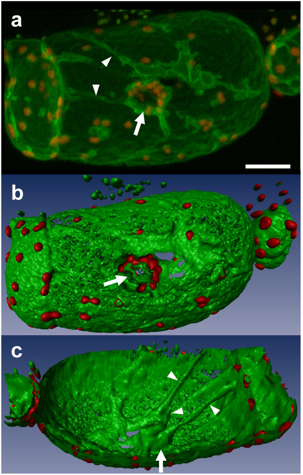
The vegetative plant vacuole occupies >90% of the volume in mature plant cells. Vacuoles play fundamental roles in adjusting cellular homeostasis and allowing cell growth. The composition of the vacuole and the regulation of its volume depend on the coordinated activities of the transporters and channels localized in the membrane (named tonoplast) surrounding the vacuole. While the tonoplast protein complexes are well studied, the tonoplast itself is less well described. To extend our knowledge of how the vacuole folds inside the plant cell, we present three-dimensional reconstructions of vacuoles from tobacco suspension cells expressing the tonoplast aquaporin fusion gene BobTIP26-1::gfp.
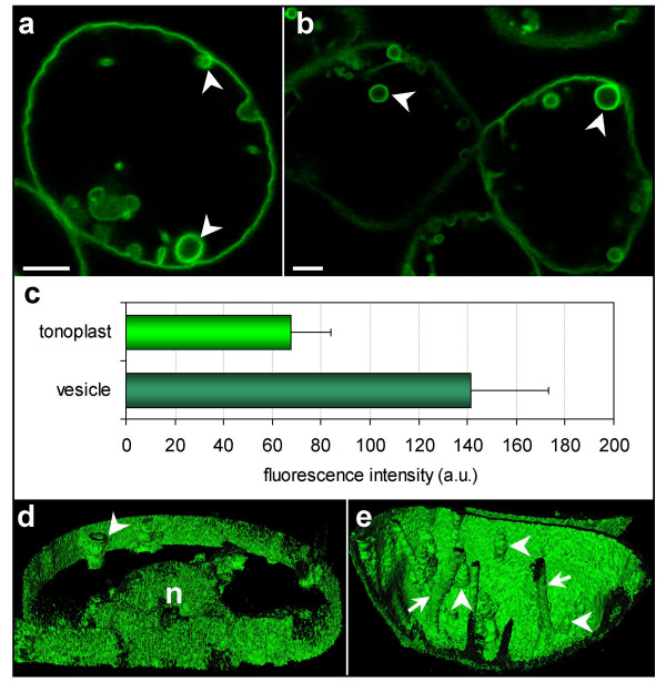
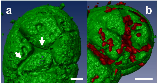
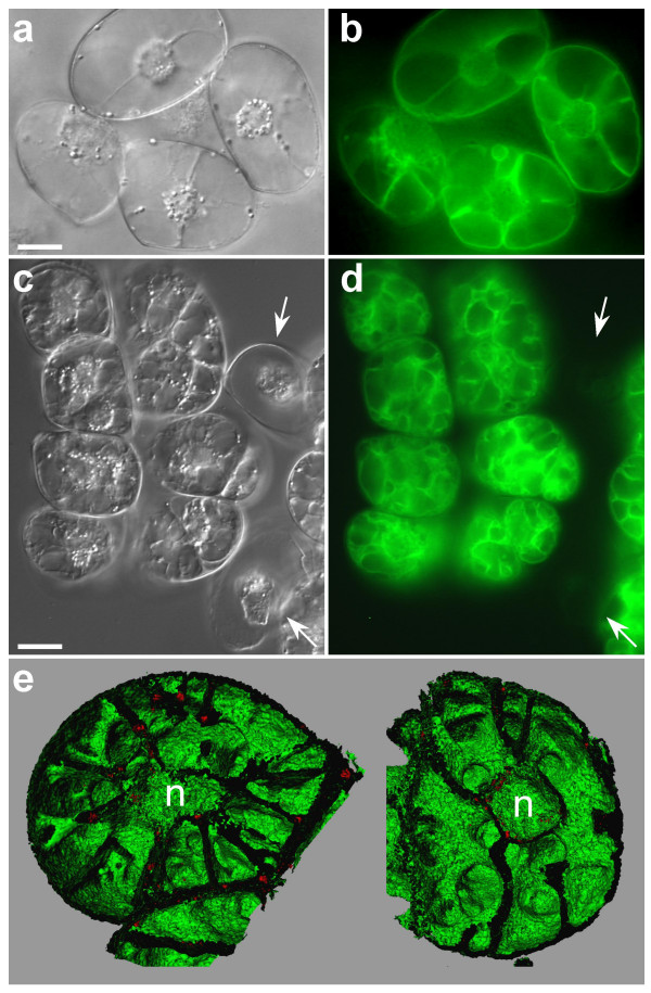
3-D reconstruction of the cell vacuole made possible an accurate analysis of large spanning folds of the vacuolar membrane under both normal and stressed conditions, and suggested interactions between surrounding plastids. Dynamic, high resolution 3-D pictures of the vacuole in tobacco suspension cells monitored under different growth conditions provide additional details about vacuolar architecture. The GFP-decorated vacuole is a single continuous compartment transected by tubular-like transvacuolar strands and large membrane surfaces. Cell culture under osmotic stress led to a complex vacuolar network with an increased tonoplast surface area. In-depth 3-D realistic inspections showed that the unity of the vacuole is maintained during acclimation to osmotic stress. Vacuolar unity exhibited during stress adaptation, coupled with the intimate associations of vacuoles with other organelles, suggests a physiological role for the vacuole in metabolism, and communication between the vacuole and organelles, respectively, in plant cells. Desiccation stress ensuing from PEG treatment generates "double" membrane structures closely linked to the tonoplast within the vacuole. These membrane structures may serve as membrane reservoirs for membrane reversion when cells are reintroduced to normal growth conditions.
3-D processing of a GFP-labeled tonoplast provides compelling visual constructions of the plant cell vacuole and elaborates on the nature of tonoplast folding and architecture. Furthermore, these methods allow real-time determination of membrane rearrangements during stresses.
植物营养液泡在成熟植物细胞中所占体积超过90%。液泡在调节细胞稳态和促进细胞生长方面发挥着重要作用。液泡的组成及其体积调节取决于位于液泡周围膜(称为液泡膜)上的转运蛋白和通道的协同活动。虽然液泡膜蛋白复合物已得到充分研究,但液泡膜本身的描述较少。为了扩展我们对液泡在植物细胞内如何折叠的认识,我们展示了来自表达液泡膜水通道蛋白融合基因BobTIP26-1::gfp的烟草悬浮细胞的液泡三维重建。
细胞液泡的三维重建使得在正常和胁迫条件下对液泡膜的大跨度折叠进行准确分析成为可能,并提示了周围质体之间的相互作用。在不同生长条件下监测的烟草悬浮细胞中液泡的动态、高分辨率三维图像提供了关于液泡结构的更多细节。用绿色荧光蛋白标记的液泡是一个单一的连续区室,被管状的跨液泡链和大的膜表面横切。渗透胁迫下的细胞培养导致了一个复杂的液泡网络,液泡膜表面积增加。深入的三维真实观察表明,在适应渗透胁迫过程中液泡的整体性得以维持。胁迫适应过程中表现出的液泡整体性,以及液泡与其他细胞器的紧密联系,分别暗示了液泡在植物细胞代谢中的生理作用以及液泡与细胞器之间的通讯。聚乙二醇处理引起的干燥胁迫在液泡内产生与液泡膜紧密相连的“双层”膜结构。当细胞重新回到正常生长条件时,这些膜结构可能作为膜储备用于膜的恢复。
对绿色荧光蛋白标记的液泡膜进行三维处理提供了令人信服的植物细胞液泡视觉构建,并阐述了液泡膜折叠和结构的本质。此外,这些方法允许实时确定胁迫期间的膜重排。