Radtke Christine, Akiyama Yukinori, Lankford Karen L, Vogt Peter M, Krause Diane S, Kocsis Jeffery D
Department of Neurology, LCI 7, P.O. Box 208018, Yale University School of Medicine, New Haven, CT 06516, USA.
Neurosci Lett. 2005 Oct 21;387(2):85-9. doi: 10.1016/j.neulet.2005.06.073.
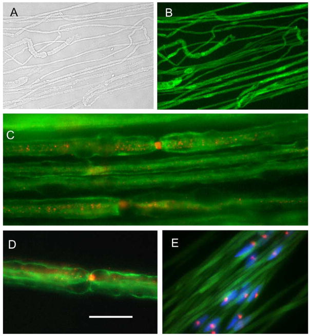
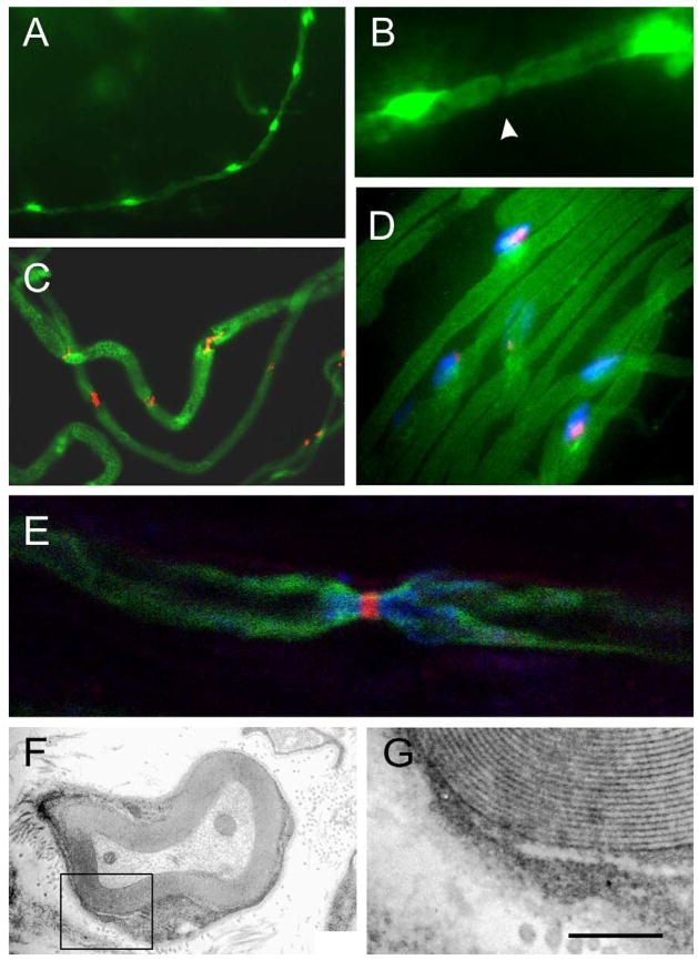
Transplantation of myelin-forming cells can remyelinate axons, but little is known of the sodium channel organization of axons myelinated by donor cells. Sciatic nerve axons of female wild type mice were transected by a crush injury and Schwann cells (SCs) from green fluorescence protein (GFP)-expressing male mice were transplanted adjacent to the crush site. The male donor cells were identified by GFP fluorescence and fluorescence in situ hybridization (FISH) for Y chromosome. In nerves of GFP-expressing mice, GFP was observed in the axoplasm and in the cytoplasmic compartments of the Schwann cells, but not in the myelin. Following transplantation of GFP-SCs into crushed nerve of wild type mice, immuno-electron microscopic analysis indicated that GFP was observed in the cytoplasmic compartments of engrafted Schwann cells which formed myelin. Nodal and paranodal regions of the axons myelinated by the GFP-SCs were identified by Na(v)1.6 sodium channel and Caspr immunostaining, respectively. Nuclear identification of the Y chromosome by FISH confirmed the donor origin of the myelin-forming cells. These results indicate that engrafted GFP-SCs participate in myelination of regenerated peripheral nerve fibers and that Na(v)1.6 sodium channel, which is the dominant sodium channel at normal nodes, is reconstituted on the regenerated axons.
髓鞘形成细胞的移植可以使轴突重新髓鞘化,但对于由供体细胞髓鞘化的轴突的钠通道组织却知之甚少。雌性野生型小鼠的坐骨神经轴突因挤压伤而横断,并将来自表达绿色荧光蛋白(GFP)的雄性小鼠的雪旺细胞(SCs)移植到挤压部位附近。通过GFP荧光和Y染色体的荧光原位杂交(FISH)鉴定雄性供体细胞。在表达GFP的小鼠的神经中,在轴质和雪旺细胞的细胞质区室中观察到GFP,但在髓鞘中未观察到。将GFP-SCs移植到野生型小鼠的挤压神经中后,免疫电子显微镜分析表明,在形成髓鞘的植入雪旺细胞的细胞质区室中观察到GFP。分别通过Na(v)1.6钠通道和Caspr免疫染色鉴定由GFP-SCs髓鞘化的轴突的结区和旁结区。通过FISH对Y染色体进行核鉴定证实了髓鞘形成细胞的供体来源。这些结果表明,植入的GFP-SCs参与再生外周神经纤维的髓鞘形成,并且正常结处的主要钠通道Na(v)1.6钠通道在再生轴突上得以重建。