Taylor Anne M, Blurton-Jones Mathew, Rhee Seog Woo, Cribbs David H, Cotman Carl W, Jeon Noo Li
Department of Biomedical Engineering, University of California, Irvine, 204 Rockwell Engineering, Irvine, California 92697, USA.
Nat Methods. 2005 Aug;2(8):599-605. doi: 10.1038/nmeth777.
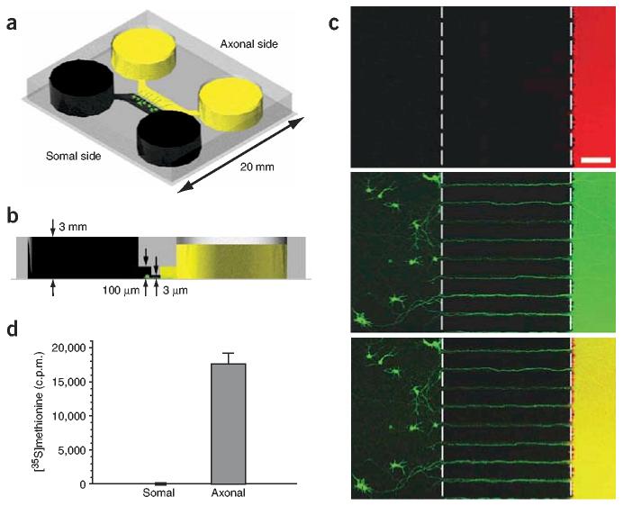
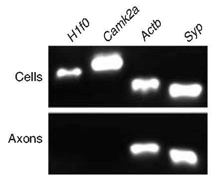
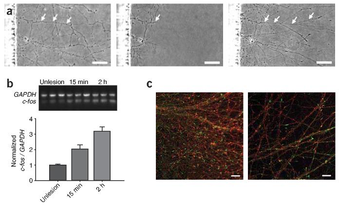
Investigation of axonal biology in the central nervous system (CNS) is hindered by a lack of an appropriate in vitro method to probe axons independently from cell bodies. Here we describe a microfluidic culture platform that polarizes the growth of CNS axons into a fluidically isolated environment without the use of targeting neurotrophins. In addition to its compatibility with live cell imaging, the platform can be used to (i) isolate CNS axons without somata or dendrites, facilitating biochemical analyses of pure axonal fractions and (ii) localize physical and chemical treatments to axons or somata. We report the first evidence that presynaptic (Syp) but not postsynaptic (Camk2a) mRNA is localized to developing rat cortical and hippocampal axons. The platform also serves as a straightforward, reproducible method to model CNS axonal injury and regeneration. The results presented here demonstrate several experimental paradigms using the microfluidic platform, which can greatly facilitate future studies in axonal biology.
中枢神经系统(CNS)轴突生物学的研究受到阻碍,因为缺乏一种合适的体外方法来独立于细胞体探测轴突。在此,我们描述了一种微流控培养平台,该平台可在不使用靶向神经营养因子的情况下,将中枢神经系统轴突的生长极化到一个流体隔离的环境中。除了与活细胞成像兼容外,该平台还可用于:(i)分离没有胞体或树突的中枢神经系统轴突,便于对纯轴突部分进行生化分析;(ii)将物理和化学处理定位到轴突或胞体。我们报告了第一个证据,即突触前(Syp)而非突触后(Camk2a)mRNA定位于发育中的大鼠皮质和海马轴突。该平台还提供了一种简单、可重复的方法来模拟中枢神经系统轴突损伤和再生。本文展示的结果证明了使用微流控平台的几种实验范式,这可极大地促进未来轴突生物学的研究。