Kielczewski Jennifer L, Pease Mary Ellen, Quigley Harry A
Glaucoma Research Laboratory, Wilmer Eye Institute, Johns Hopkins University School of Medicine, 600 North Wolfe Street, Baltimore, MD, USA.
Invest Ophthalmol Vis Sci. 2005 Sep;46(9):3188-96. doi: 10.1167/iovs.05-0321.
To detect alterations in amacrine cells associated with retinal ganglion cell (RGC) depletion caused by experimental optic nerve transection and glaucoma.
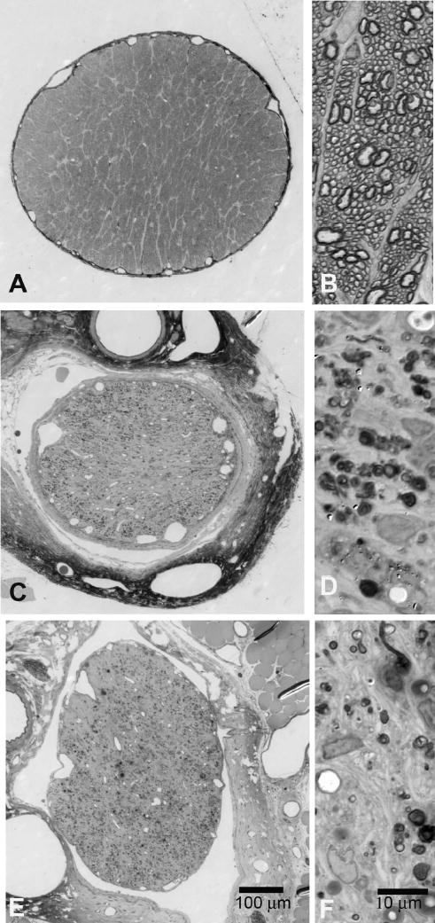
Intraocular pressure (IOP) was elevated unilaterally in 18 rats by translimbal trabecular laser treatment, and eyes were studied at 1 (n = 6), 2 (n = 5), and 3 (n = 7) months. Complete optic nerve transection was performed unilaterally in nine rats with survival for 1 (n = 4) and 3 (n = 5) months. Serial cryosections (five per eye) were immunohistochemically labeled with rabbit anti-gamma-aminobutyric acid (GABA) and anti-glycine antibodies. Cells in the ganglion cell and inner nuclear layers that labeled for GABA or glycine were counted in a masked fashion under bright-field microscopy. Additional labeling with other RGC and amacrine antigens was also performed. RGC loss was quantified by axon counts.
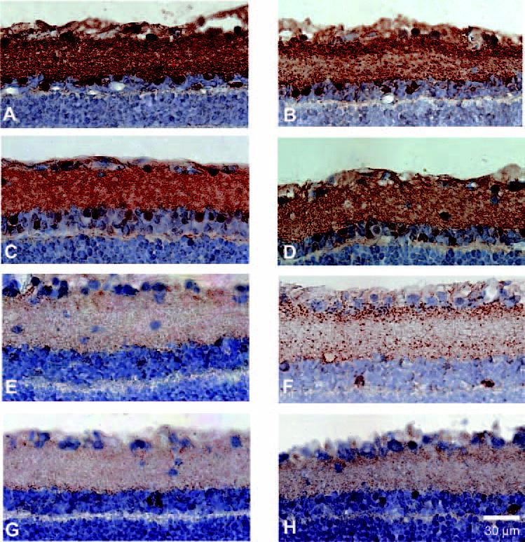
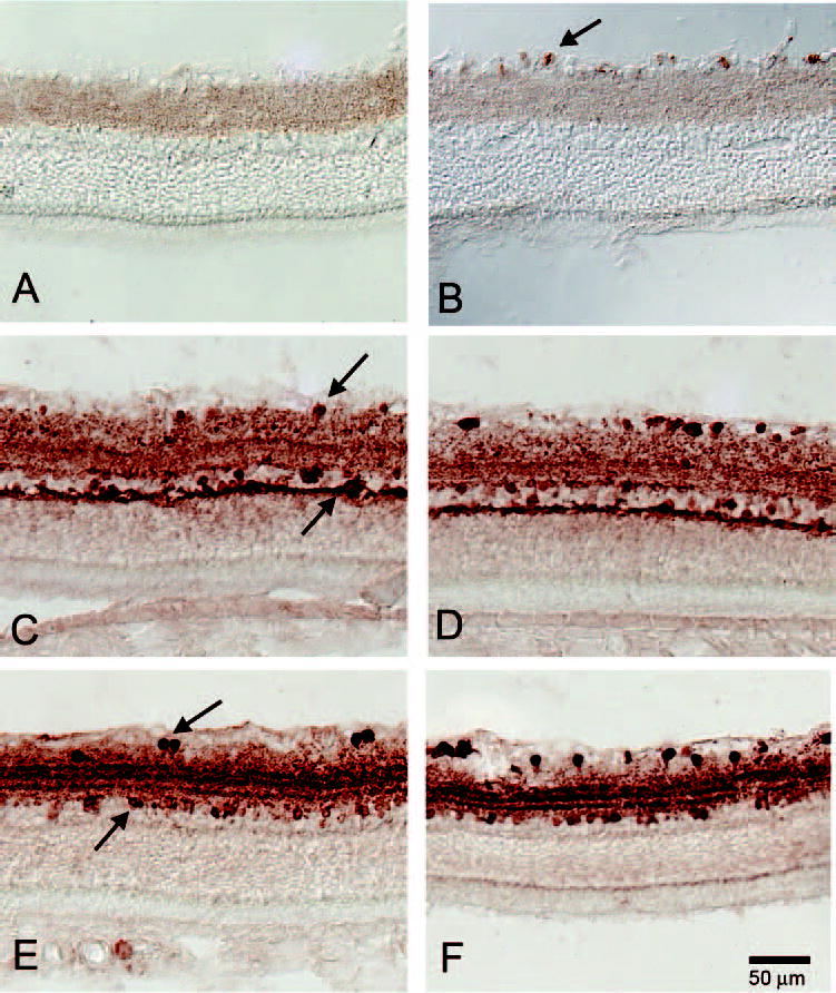
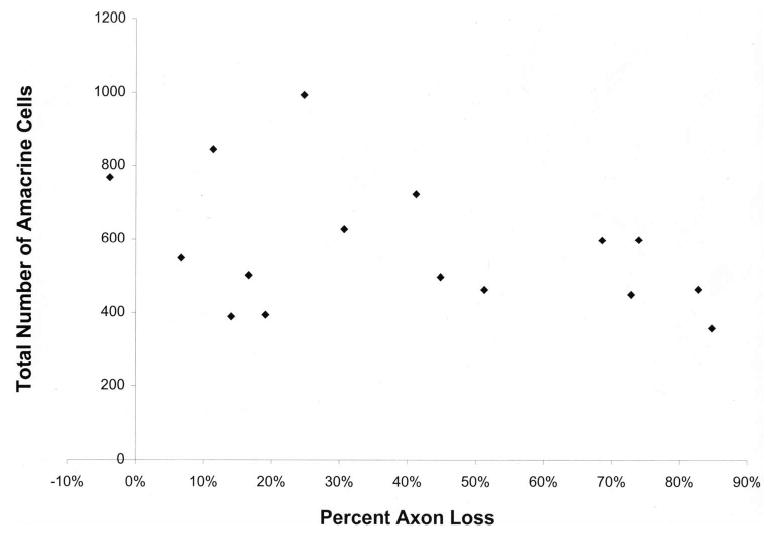
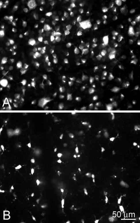
Amacrine cells identified by GABA and glycine labeling were not significantly affected by experimental glaucoma, with a mean decrease of 15% compared with bilaterally untreated control cells (557 +/- 186 neurons/mm [glaucoma] versus 653.9 +/- 114.4 neurons/mm [control] of retina; P = 0.15, t-test). There was no significant trend for amacrine cell counts to be lower in eyes with fewer RGCs (r = -0.39, P = 0.11). By contrast, there was highly significant loss of GABA and glycine staining 3 months after nerve transection, both in the treated and the fellow eyes (P < 0.0001, t-test). However, there was a substantial number of remaining amacrine cells in transected retinas, as indicated by labeling for calretinin and calbindin.
Experimental glaucoma causes minimal change in amacrine cells and their expression of neurotransmitters. After nerve transection, neurotransmitter presence declines, but many amacrine cell bodies remain. Differences among optic nerve injury models, as well as effects on "untreated" fellow eyes, should be recognized.
检测与实验性视神经横断和青光眼所致视网膜神经节细胞(RGC)缺失相关的无长突细胞的改变。
通过经角膜小梁激光治疗使18只大鼠单侧眼压升高,并在1个月(n = 6)、2个月(n = 5)和3个月(n = 7)时对眼睛进行研究。对9只大鼠进行单侧完全性视神经横断,分别存活1个月(n = 4)和3个月(n = 5)。连续冰冻切片(每只眼5张)用兔抗γ-氨基丁酸(GABA)和抗甘氨酸抗体进行免疫组织化学标记。在明场显微镜下以盲法计数神经节细胞层和内核层中标记GABA或甘氨酸的细胞。还进行了其他RGC和无长突细胞抗原的额外标记。通过轴突计数对RGC缺失进行量化。
通过GABA和甘氨酸标记鉴定的无长突细胞受实验性青光眼的影响不显著,与双侧未治疗的对照细胞相比平均减少15%(视网膜青光眼组为557±186个神经元/mm,对照组为653.9±114.4个神经元/mm;P = 0.15,t检验)。在RGC较少的眼中,无长突细胞计数没有显著降低的趋势(r = -0.39,P = 0.11)。相比之下,神经横断3个月后,无论是治疗眼还是对侧眼,GABA和甘氨酸染色均有高度显著的缺失(P < 0.0001,t检验)。然而,如钙视网膜蛋白和钙结合蛋白标记所示,横断视网膜中仍有大量剩余的无长突细胞。
实验性青光眼导致无长突细胞及其神经递质表达的变化极小。神经横断后,神经递质的存在减少,但许多无长突细胞体仍然存在。应认识到视神经损伤模型之间的差异以及对“未治疗”对侧眼的影响。