Peterson James J, Orisme Wilda, Fellows Jonathan, McDowell J Hugh, Shelamer Charles L, Dugger Donald R, Smith W Clay
Department of Ophthalmology, University of Florida, Gainesville, 32610, USA.
Invest Ophthalmol Vis Sci. 2005 Nov;46(11):3988-98. doi: 10.1167/iovs.05-0567.
Light-driven protein translocation is responsible for the dramatic redistribution of some proteins in vertebrate rod photoreceptors. In this study, the involvement of microtubules and microfilaments in the light-driven translocation of arrestin and transducin was investigated.
Pharmacologic reagents were applied to native and transgenic Xenopus tadpoles, to disrupt the microtubules (thiabendazole) and microfilaments (cytochalasin D and latrunculin B) of the rod photoreceptors. Quantitative confocal imaging was used to assess the impact of these treatments on arrestin and transducin translocation. A series of transgenic tadpoles expressing arrestin truncations were also created to identify portions of arrestin that enable arrestin to translocate.
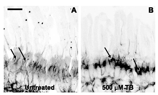
Application of cytochalasin D or latrunculin B to disrupt the microfilament organization selectively slowed only transducin movement from the inner to the outer segments. Perturbation of the microtubule cytoskeleton with thiabendazole slowed the translocation of both arrestin and transducin, but only in moving from the outer to the inner segments. Transgenic Xenopus expressing fusions of green fluorescent protein (GFP) with portions of arrestin implicates the C terminus of arrestin as an important portion of the molecule for promoting translocation. This C-terminal region can be used independently to promote translocation of GFP in response to light.
The results show that disruption of the cytoskeletal network in rod photoreceptors has specific effects on the translocation of arrestin and transducin. These effects suggest that the light-driven translocation of visual proteins at least partially relies on an active motor-driven mechanism for complete movement of arrestin and transducin.
光驱动的蛋白质转位负责脊椎动物视杆光感受器中某些蛋白质的显著重新分布。在本研究中,研究了微管和微丝在视紫红质抑制蛋白和转导蛋白的光驱动转位中的作用。
将药理试剂应用于天然和转基因非洲爪蟾蝌蚪,以破坏视杆光感受器的微管(噻苯咪唑)和微丝(细胞松弛素D和拉特罗毒素B)。使用定量共聚焦成像来评估这些处理对视紫红质抑制蛋白和转导蛋白转位的影响。还创建了一系列表达视紫红质抑制蛋白截短体的转基因蝌蚪,以鉴定使视紫红质抑制蛋白能够转位的部分。
应用细胞松弛素D或拉特罗毒素B破坏微丝组织仅选择性地减缓了转导蛋白从内段向外段的移动。用噻苯咪唑扰乱微管细胞骨架减缓了视紫红质抑制蛋白和转导蛋白的转位,但仅在从外段向内段移动时。表达绿色荧光蛋白(GFP)与视紫红质抑制蛋白部分融合的转基因非洲爪蟾表明视紫红质抑制蛋白的C末端是促进转位的分子的重要部分。该C末端区域可独立用于促进GFP响应光的转位。
结果表明,视杆光感受器中细胞骨架网络的破坏对视紫红质抑制蛋白和转导蛋白的转位有特定影响。这些影响表明,视觉蛋白的光驱动转位至少部分依赖于一种主动的马达驱动机制,以实现视紫红质抑制蛋白和转导蛋白的完全移动。