Maker Ajay V, Attia Peter, Rosenberg Steven A
Surgery Branch, National Cancer Institute, National Institutes of Health, Bethesda, MD 20814, USA.
J Immunol. 2005 Dec 1;175(11):7746-54. doi: 10.4049/jimmunol.175.11.7746.
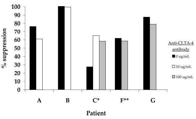
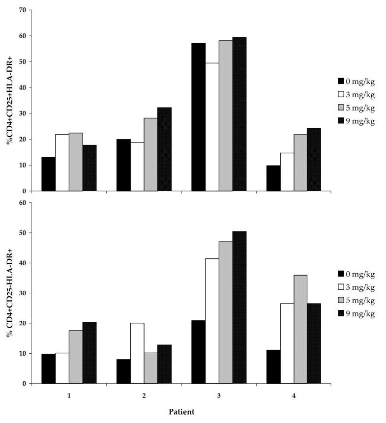
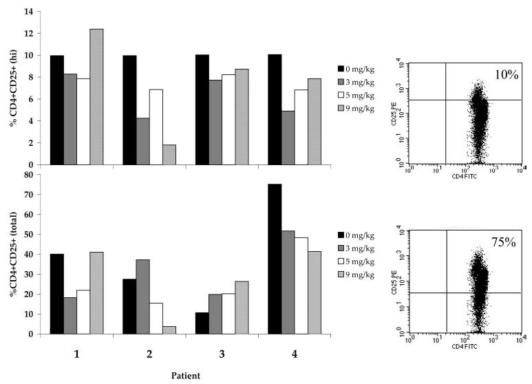
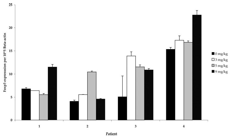
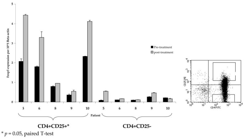
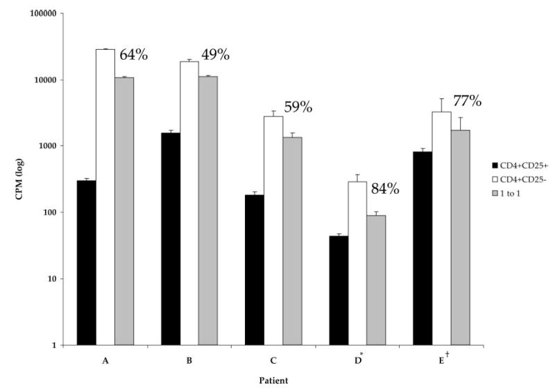
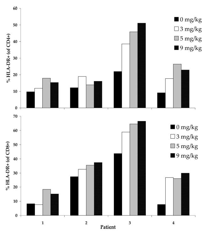
We have demonstrated previously that the administration of CTLA-4 blockade has mediated objective cancer regression and autoimmunity in patients with metastatic melanoma. To explore the mechanism of these in vivo effects, we have studied the changes in lymphocyte phenotype and function in patients receiving anti-CTLA-4 Ab (MDX-010). Patients with stage IV melanoma or renal cell cancer were treated every 3 wk with an anti-CTLA-4 Ab with or without peptide immunization. Pheresis samples were analyzed using flow cytometry to determine lymphocyte cell surface markers. Gene expression analyses and proliferation assays were conducted on purified T cell subsets. Anti-CTLA-4 Ab did not inhibit the suppressive activity of CD4+CD25+ cells in vitro or in vivo. In addition, there was no decrease in the expression of CD4+CD25+ cells in whole PBMC, nor a decrease in Foxp3 gene expression in the CD4+ or CD4+CD25+ purified cell populations posttreatment. The percentage of CD4+, CD8+, CD4+CD25+, and CD4+CD25- T cells in PBMC expressing the activation marker HLA-DR increased following anti-CTLA-4 Ab administration. Therefore, our results suggest that the antitumor effects of CTLA-4 blockade are due to increased T cell activation rather than inhibition or depletion of T regulatory cells.
我们之前已经证明,给予CTLA-4阻断剂可介导转移性黑色素瘤患者的客观癌症消退和自身免疫。为了探究这些体内效应的机制,我们研究了接受抗CTLA-4抗体(MDX-010)治疗的患者淋巴细胞表型和功能的变化。IV期黑色素瘤或肾细胞癌患者每3周接受一次抗CTLA-4抗体治疗,同时或不同时进行肽免疫。使用流式细胞术分析血细胞分离样本以确定淋巴细胞细胞表面标志物。对纯化的T细胞亚群进行基因表达分析和增殖测定。抗CTLA-4抗体在体外或体内均未抑制CD4 + CD25 +细胞的抑制活性。此外,治疗后全外周血单个核细胞(PBMC)中CD4 + CD25 +细胞的表达没有降低,CD4 +或CD4 + CD25 +纯化细胞群体中Foxp3基因表达也没有降低。给予抗CTLA-4抗体后,PBMC中表达激活标志物HLA-DR的CD4 +、CD8 +、CD4 + CD25 +和CD4 + CD25 - T细胞百分比增加。因此,我们的结果表明,CTLA-4阻断的抗肿瘤作用是由于T细胞活化增加而非调节性T细胞的抑制或耗竭。