Ramasamy Lakshminarayanan, Sperelakis Nicholas
Dept. of Molecular & Cellular Physiology, University of Cincinnati College of Medicine, Cincinnati, OH 45267-0576, USA.
Theor Biol Med Model. 2005 Dec 12;2:48. doi: 10.1186/1742-4682-2-48.
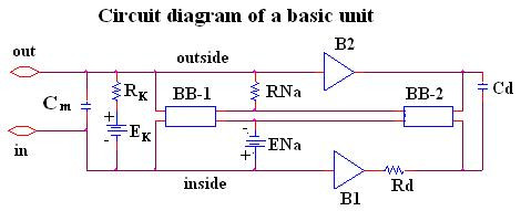
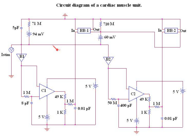
In previous studies on propagation of simulated action potentials (APs) in cardiac muscle using PSpice modeling, we reported that a second black-box (BB) could not be inserted into the K+ leg of the basic membrane unit because that caused the PSpice program to become very unstable. Therefore, only the rising phase of the APs could be simulated. This restriction was acceptable since only the mechanism of transmission of excitation from one cell to the next was being investigated.
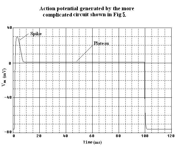
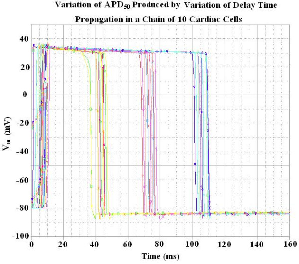
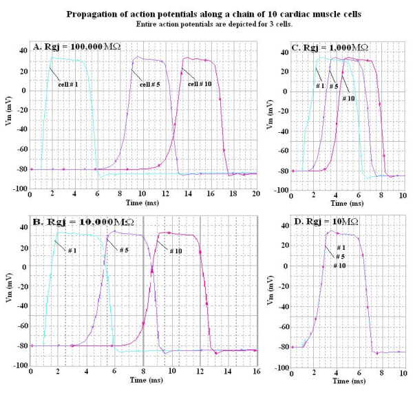
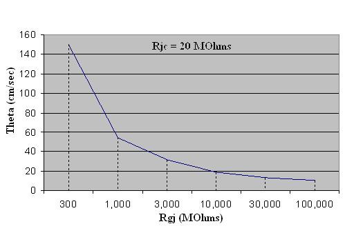
We have now been able to repolarize the AP by inserting a second BB into the Na+ leg of the basic units. This second BB effectively mimicked deactivation of the Na+ channel conductance. This produced repolarization of the AP, not by activation of K+ conductance, but by deactivation of the Na+ conductance. The propagation of complete APs was studied in a chain (strand) of 10 cardiac muscle cells, in which various numbers of gap-junction (gj) channels (assumed to be 100 pS each) were inserted across the cell junctions. The shunt resistance across the junctions produced by the gj-channels (Rgj) was varied from 100,000 M? (0 gj-channels) to 10,000 M? (1 gj-channel), to 1,000 M? (10 channels), to 100 M? (100 channels), and 10 M? (1000 channels). The velocity of propagation (theta, in cm/s) was calculated from the measured total propagation time (TPT, the time difference between when the AP rising phase of the first cell and the last cell crossed -20 mV, assuming a cell length of 150 microm. When there were no gj-channels, or only a few, the transmission of excitation between cells was produced by the electric field (EF), i.e. the negative junctional cleft potential, that is generated in the narrow junctional clefts (e.g. 100 A) when the prejunctional membrane fires an AP. When there were many gj-channels (e.g. 1000 or 10,000), the transmission of excitation was produced by local-circuit current flow from one cell to the next through the gj-channels.
We have now been able to simulate complete APs in cardiac muscle cells that could propagate along a single chain of 10 cells, even when there were no gj-channels between the cells.
在先前使用PSpice建模对心肌中模拟动作电位(AP)传播的研究中,我们报告称不能在基本膜单元的K⁺支路中插入第二个黑箱(BB),因为这会使PSpice程序变得非常不稳定。因此,只能模拟动作电位的上升阶段。由于当时仅研究兴奋从一个细胞传递到下一个细胞的机制,所以这种限制是可以接受的。
我们现在已经能够通过在基本单元的Na⁺支路中插入第二个BB使动作电位复极化。这个第二个BB有效地模拟了Na⁺通道电导的失活。这导致动作电位复极化,不是通过激活K⁺电导,而是通过使Na⁺电导失活。在由10个心肌细胞组成的链(束)中研究了完整动作电位的传播,其中在细胞连接处插入了不同数量的间隙连接(gj)通道(假设每个通道为100 pS)。由gj通道产生的跨连接处的分流电阻(Rgj)从100,000 MΩ(0个gj通道)变化到10,000 MΩ(1个gj通道)、1,000 MΩ(10个通道)、100 MΩ(100个通道)和10 MΩ(1000个通道)。传播速度(θ,单位为cm/s)根据测量的总传播时间(TPT,即第一个细胞和最后一个细胞的动作电位上升阶段越过-20 mV时的时间差,假设细胞长度为150微米)计算得出。当没有gj通道或只有少数gj通道时,细胞间的兴奋传递是由电场(EF)产生的,即当突触前膜发放动作电位时在狭窄的连接间隙(例如100 Å)中产生的负连接间隙电位。当有许多gj通道(例如1000个或10,000个)时,兴奋传递是通过局部电路电流从一个细胞通过gj通道流向下一个细胞产生的。
我们现在已经能够在心肌细胞中模拟完整的动作电位,这些动作电位能够沿着由10个细胞组成的单链传播,即使细胞之间没有gj通道。