Sperelakis Nicholas, Ramasamy Lakshminarayanan, Kalloor Bijoy
Dept. of Molecular & Cellular Physiology University of Cincinnati College of Medicine Cincinnati, OH 45267-0576 USA.
Theor Biol Med Model. 2005 Feb 14;2:5. doi: 10.1186/1742-4682-2-5.
Propagation of repolarization is a phenomenon that occurs in cardiac muscle. We wanted to test whether this phenomenon would also occur in our model of simulated action potentials (APs) of cardiac muscle (CM) and smooth muscle (SM) generated with the PSpice program.
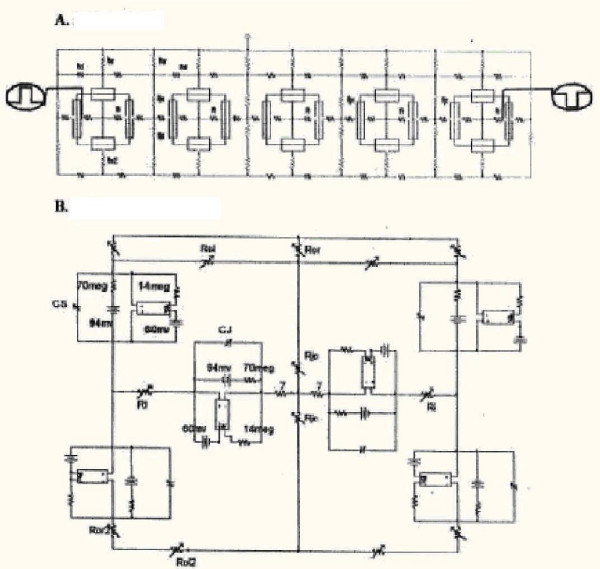
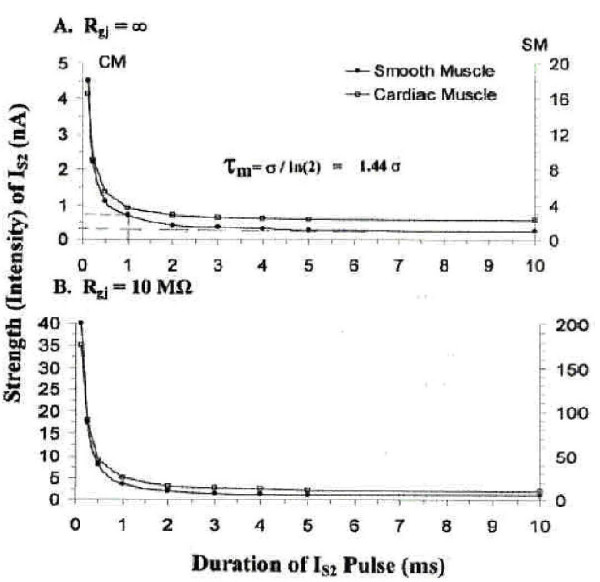
A linear chain of 5 cells was used, with intracellular stimulation of cell #1 for the antegrade propagation and of cell #5 for the retrograde propagation. The hyperpolarizing stimulus parameters applied for termination of the AP in cell #5 were varied over a wide range in order to generate strength / duration (S/D) curves. Because it was not possible to insert a second "black box" (voltage-controlled current source) into the basic units representing segments of excitable membrane that would allow the cells to respond to small hyperpolarizing voltages, gap-junction (g.j.) channels had to be inserted between the cells, represented by inserting a resistor (Rgj) across the four cell junctions.
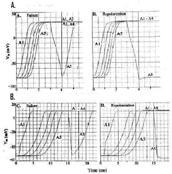
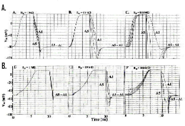
Application of sufficient hyperpolarizing current to cell #5 to bring its membrane potential (Vm) to within the range of the sigmoidal curve of the Na+ conductance (CM) or Ca++ conductance (SM) terminated the AP in cell #5 in an all-or-none fashion. If there were no g.j. channels (Rgj = infinity), then only cell #5 repolarized to its stable resting potential (RP; -80 mV for CM and -55 mV for SM). The positive junctional cleft potential (VJC) produced only a small hyperpolarization of cell #4. However, if many g.j. channels were inserted, more hyperpolarizing current was required (for a constant duration) to repolarize cell #5, but repolarization then propagated into cells 4, 3, 2, and 1. When duration of the pulses was varied, a typical S/D curve, characteristic of excitable membranes, was produced. The chronaxie measured from the S/D curve was about 1.0 ms, similar to that obtained for muscle membranes.
These experiments demonstrate that normal antegrade propagation of excitation can occur in the complete absence of g.j. channels, and therefore no low-resistance pathways between cells, by the electric field (negative VJC) developed in the narrow junctional clefts. Because it was not possible to insert a second black-box into the basic units that would allow the cells to respond to small hyperpolarizing voltages, only cell #5 (the cell injected with hyperpolarizing pulses) repolarized in an all-or-none manner. But addition of many g.j. channels allowed repolarization to propagate in a retrograde direction over all 5 cells.
复极化传播是一种发生在心肌中的现象。我们想要测试这种现象是否也会在我们用PSpice程序生成的心肌(CM)和平滑肌(SM)模拟动作电位(AP)模型中出现。
使用由5个细胞组成的线性链,对细胞#1进行细胞内刺激以实现顺行传播,对细胞#5进行细胞内刺激以实现逆行传播。为了生成强度/持续时间(S/D)曲线,在很宽的范围内改变施加于细胞#5以终止AP的超极化刺激参数。由于不可能在代表可兴奋膜段的基本单元中插入第二个“黑匣子”(电压控制电流源)以使细胞能够响应小的超极化电压,因此必须在细胞之间插入间隙连接(g.j.)通道,这通过在四个细胞连接处跨接一个电阻器(Rgj)来表示。
向细胞#5施加足够的超极化电流,使其膜电位(Vm)处于钠电导(CM)或钙电导(SM)的S形曲线范围内,以全或无的方式终止细胞#5中的AP。如果没有g.j.通道(Rgj =无穷大),那么只有细胞#5复极化到其稳定的静息电位(RP;CM为 -80 mV,SM为 -55 mV)。正向连接间隙电位(VJC)仅使细胞#4产生小的超极化。然而,如果插入许多g.j.通道,则需要更多的超极化电流(在恒定持续时间内)使细胞#5复极化,但随后复极化会传播到细胞4、3、2和1。当改变脉冲持续时间时,会产生一条典型的、具有可兴奋膜特征的S/D曲线。从S/D曲线测得的时值约为1.0 ms,与肌肉膜的时值相似。
这些实验表明,在完全没有g.j.通道(即细胞间没有低电阻通路)的情况下,通过狭窄连接间隙中产生的电场(负VJC),兴奋的正常顺行传播仍可发生。由于不可能在基本单元中插入第二个黑匣子以使细胞能够响应小的超极化电压,所以只有细胞#5(注入超极化脉冲的细胞)以全或无的方式复极化。但是添加许多g.j.通道后,复极化能够在所有5个细胞中逆行传播。