Kucera Jan P, Rohr Stephan, Rudy Yoram
Department of Physiology, University of Bern, Switzerland.
Circ Res. 2002 Dec 13;91(12):1176-82. doi: 10.1161/01.res.0000046237.54156.0a.
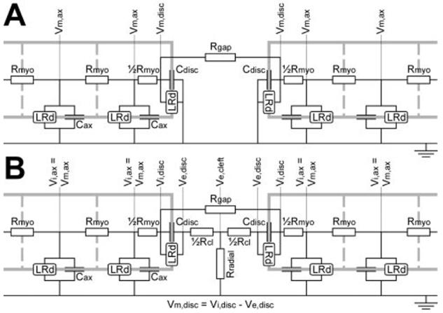
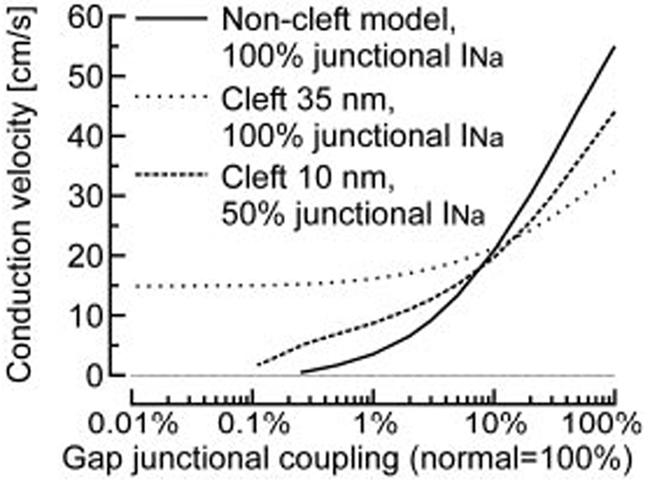
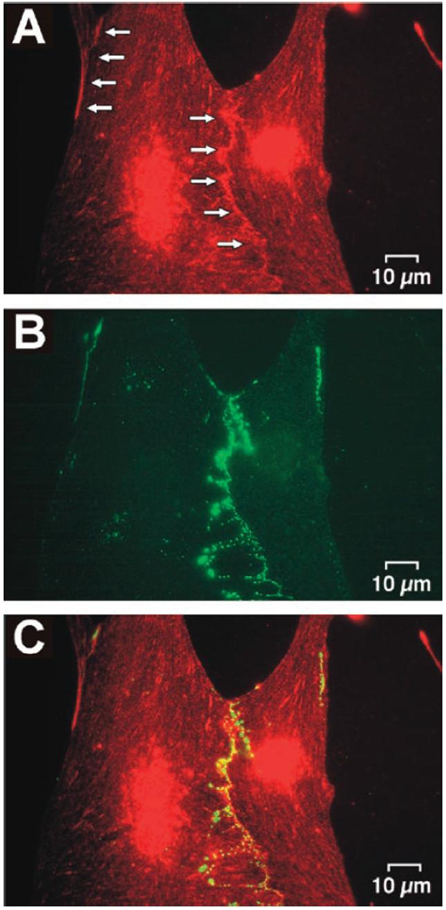
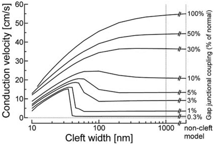
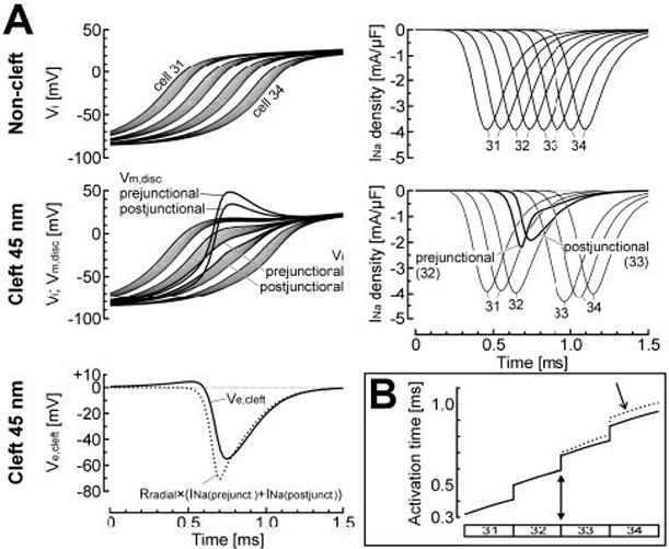
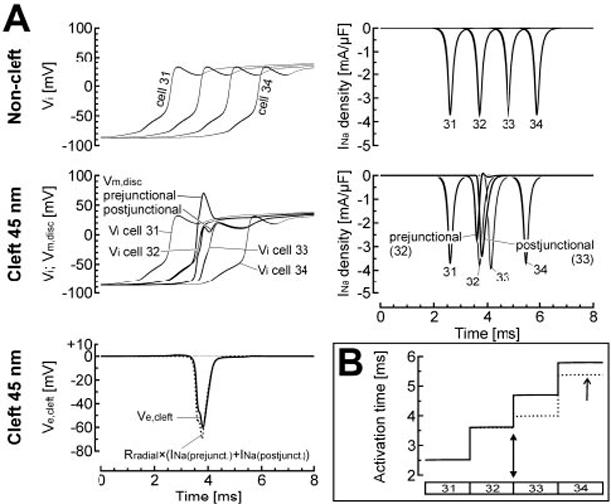
It is well known that the sodium current (I(Na)) and the degree of gap-junctional electrical coupling are the key determinants of action potential (AP) conduction in cardiac tissue. Immunohistochemical studies have shown that sodium channels (NaChs) are preferentially located in intercalated disks (IDs). Using dual immunocytochemical staining, we confirmed the colocalization of NaChs with connexin43 in cultures of neonatal rat ventricular myocytes. In mathematical simulations of conduction using the Luo-Rudy dynamic model of the ventricular AP, we assessed the hypothesis that conduction could be modulated by the preferential localization of NaChs in IDs. Localization of I(Na) at the ID caused a large negative potential in the intercellular cleft, which influenced conduction in two opposing ways, depending on the degree of electrical coupling: (1) for normal and moderately reduced coupling, the negative cleft potential led to a large overshoot of the transmembrane potential resulting in a decreased driving force for I(Na) itself (self-attenuation), which slowed conduction; (2) for greatly reduced coupling (<10%), the negative cleft potential induced by I(Na) in the prejunctional membrane led to suprathreshold depolarization of the postjunctional membrane, which facilitated and accelerated conduction. When cleft potential effects were not incorporated, conduction was not significantly affected by the ID localization of I(Na). By enhancing conduction through the establishment of cleft potentials, the localization of NaChs in IDs might protect the myocardium from conduction block, very slow conduction, and microreentry under conditions of greatly reduced coupling. Conversely, by supporting moderately slow conduction, this mechanism could also promote arrhythmias.
众所周知,钠电流(I(Na))和缝隙连接电偶联程度是心脏组织中动作电位(AP)传导的关键决定因素。免疫组织化学研究表明,钠通道(NaChs)优先定位于闰盘(IDs)。通过双重免疫细胞化学染色,我们在新生大鼠心室肌细胞培养物中证实了NaChs与连接蛋白43的共定位。在使用心室AP的Luo-Rudy动态模型进行传导的数学模拟中,我们评估了NaChs在IDs中的优先定位可调节传导的假设。I(Na)在ID处的定位导致细胞间裂隙中出现大的负电位,这以两种相反的方式影响传导,具体取决于电偶联程度:(1)对于正常和适度降低的偶联,负的裂隙电位导致跨膜电位大幅超射,导致I(Na)自身的驱动力降低(自我衰减),从而减慢传导;(2)对于大幅降低的偶联(<10%),I(Na)在前膜中诱导的负裂隙电位导致后膜超阈值去极化,从而促进和加速传导。当不考虑裂隙电位效应时,I(Na)在ID处的定位对传导没有显著影响。通过建立裂隙电位来增强传导,NaChs在IDs中的定位可能在偶联大幅降低的情况下保护心肌免受传导阻滞、极慢传导和微折返的影响。相反,通过支持适度缓慢的传导,这种机制也可能促进心律失常。