Sleeman Katherine E, Kendrick Howard, Ashworth Alan, Isacke Clare M, Smalley Matthew J
Breakthrough Breast Cancer Research Centre, The Institute of Cancer Research, Fulham Road, London SW3 6JB, UK.
Breast Cancer Res. 2006;8(1):R7. doi: 10.1186/bcr1371. Epub 2005 Dec 12.
Breast cancer is thought to arise in mammary epithelial stem cells. There is, therefore, a large amount of interest in identifying these cells. The breast is a complex tissue consisting of two epithelial layers (an outer myoepithelial/basal layer and an inner luminal epithelial layer) as well as a large non-epithelial component (fibroblasts, endothelial cells, lymphocytes, adipocytes, neurons and myocytes). The definitive identification of a mammary epithelial stem cell population is critically dependent on its purity. To date, this has been hampered by the lack of suitable markers to separate out the two epithelial layers, and to remove contaminating non-epithelial cells.
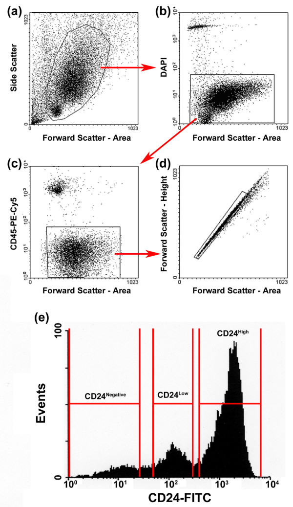
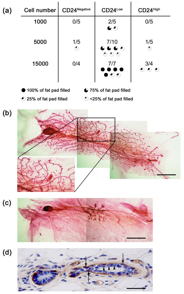
Mouse mammary glands were dissociated and stained with CD24. Cells were sorted into separate populations based on CD24 expression and assessed for luminal epithelial and myoepithelial/basal markers by direct fluorescent microscopy and real time PCR. The stem/progenitor potential of these cell populations was assessed in vivo by cleared mammary fat pad transplantation.
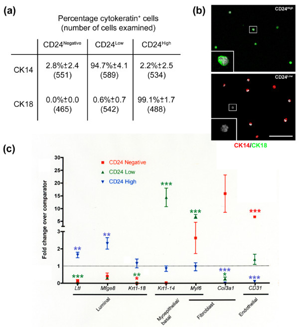
Three populations of CD24 expressing cells were identified: CD24Negative, CD24Low and CD24High. Staining of these cells with cytokeratin markers revealed that these populations correspond to non-epithelial, myoepithelial/basal and luminal epithelial cells, respectively. Cell identities were confirmed by quantitative PCR. Cleared mammary fat pad transplantation of these cell populations revealed that extensive mammary fat pad repopulation capacity segregates with the CD24Low cells, whilst CD24High cells have limited repopulation capacity.
Differential staining of mammary epithelial cells for CD24 can be used to simultaneously isolate pure populations of non-epithelial, myoepithelial/basal and luminal epithelial cells. Furthermore, mammary fat pad repopulation capacity is enriched in the CD24Low population. As separation is achieved using a single marker, it will be possible to incorporate additional markers to further subdivide these populations. This will considerably facilitate the further analysis of mammary epithelial subpopulations, whilst ensuring high purity, which is key for understanding mammary epithelial stem cells in normal tissue biology and carcinogenesis.
乳腺癌被认为起源于乳腺上皮干细胞。因此,识别这些细胞引起了广泛关注。乳腺是一个复杂的组织,由两层上皮细胞(外层的肌上皮/基底层和内层的管腔上皮层)以及大量非上皮成分(成纤维细胞、内皮细胞、淋巴细胞、脂肪细胞、神经元和肌细胞)组成。乳腺上皮干细胞群体的明确鉴定关键取决于其纯度。迄今为止,由于缺乏合适的标记物来分离这两层上皮细胞并去除污染的非上皮细胞,这一鉴定工作受到了阻碍。
将小鼠乳腺解离并用CD24染色。根据CD24表达将细胞分选到不同群体中,并通过直接荧光显微镜和实时PCR评估管腔上皮和肌上皮/基底标记物。通过清除乳腺脂肪垫移植在体内评估这些细胞群体的干细胞/祖细胞潜能。
鉴定出三个表达CD24的细胞群体:CD24阴性、CD24低表达和CD24高表达。用细胞角蛋白标记物对这些细胞进行染色显示,这些群体分别对应非上皮细胞、肌上皮/基底细胞和管腔上皮细胞。通过定量PCR确认了细胞身份。对这些细胞群体进行清除乳腺脂肪垫移植显示,广泛的乳腺脂肪垫重新填充能力与CD24低表达细胞相关,而CD24高表达细胞的重新填充能力有限。
对乳腺上皮细胞进行CD24差异染色可用于同时分离纯的非上皮细胞、肌上皮/基底细胞和管腔上皮细胞群体。此外,乳腺脂肪垫重新填充能力在CD24低表达群体中富集。由于使用单一标记物即可实现分离,因此可以加入其他标记物进一步细分这些群体。这将极大地促进对乳腺上皮亚群的进一步分析,同时确保高纯度,这对于理解正常组织生物学和致癌过程中的乳腺上皮干细胞至关重要。