Dawn Buddhadeb, Guo Yiru, Rezazadeh Arash, Huang Yiming, Stein Adam B, Hunt Greg, Tiwari Sumit, Varma Jai, Gu Yan, Prabhu Sumanth D, Kajstura Jan, Anversa Piero, Ildstad Suzanne T, Bolli Roberto
Institute of Molecular Cardiology, University of Louisville, Louisville, KY 40292, USA.
Circ Res. 2006 Apr 28;98(8):1098-105. doi: 10.1161/01.RES.0000218454.76784.66. Epub 2006 Mar 23.
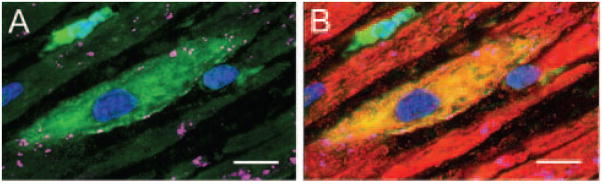
We systematically investigated the comparative efficacy of three different cytokine regimens, administered after a reperfused myocardial infarction, in regenerating cardiac tissue and improving left ventricular (LV) function. Wild-type (WT) mice underwent a 30-minute coronary occlusion followed by reperfusion and received vehicle, granulocyte colony-stimulating factor (G-CSF)+Flt-3 ligand (FL), G-CSF+stem cell factor (SCF), or G-CSF alone starting 4 hours after reperfusion. In separate experiments, chimeric mice generated by reconstitution of radioablated WT mice with bone marrow from enhanced green fluorescent protein (EGFP) transgenic mice underwent identical protocols. Mice were euthanized 5 weeks later. Echocardiographically, LV function was improved in G-CSF+FL- and G-CSF+SCF-treated but not in G-CSF-treated mice, whereas LV end-diastolic dimensions were smaller in all three groups. Morphometrically, cytokine-treated hearts had smaller LV diameter and volume. Numerous EGFP-positive cardiomyocytes, capillaries, and arterioles were noted in the infarcted region in cytokine-treated chimeric mice treated with G-CSF+FL or G-CSF+SCF, but the numbers were much smaller in G-CSF-treated mice. G-CSF+FL therapy mobilized bone marrow-derived cells exhibiting increased expression of surface antigens (CD62L and CD11a) that facilitate homing. We conclude that postinfarct cytokine therapy with G-CSF+FL or G-CSF+SCF limits adverse LV remodeling and improves LV performance by promoting cardiac regeneration and probably also by exerting other beneficial actions unrelated to regeneration, and that G-CSF alone is less effective.
我们系统地研究了三种不同细胞因子方案在再灌注心肌梗死后促进心脏组织再生和改善左心室(LV)功能方面的比较疗效。野生型(WT)小鼠经历30分钟的冠状动脉闭塞,随后进行再灌注,并在再灌注后4小时开始接受载体、粒细胞集落刺激因子(G-CSF)+Flt-3配体(FL)、G-CSF+干细胞因子(SCF)或单独的G-CSF治疗。在单独的实验中,用增强型绿色荧光蛋白(EGFP)转基因小鼠的骨髓重建经放射性消融的WT小鼠所产生的嵌合小鼠接受相同的方案。5周后对小鼠实施安乐死。超声心动图显示,接受G-CSF+FL和G-CSF+SCF治疗的小鼠左心室功能得到改善,而接受G-CSF治疗的小鼠则未改善,而三组小鼠的左心室舒张末期内径均较小。形态学测量显示,接受细胞因子治疗的心脏左心室直径和容积较小。在用G-CSF+FL或G-CSF+SCF治疗的细胞因子处理的嵌合小鼠的梗死区域中,发现了大量EGFP阳性心肌细胞、毛细血管和小动脉,但在接受G-CSF治疗的小鼠中数量要少得多。G-CSF+FL疗法动员了骨髓来源的细胞,这些细胞表面抗原(CD62L和CD11a)的表达增加,有利于归巢。我们得出结论,梗死后期用G-CSF+FL或G-CSF+SCF进行细胞因子治疗可限制不良的左心室重构,并通过促进心脏再生以及可能还通过发挥与再生无关的其他有益作用来改善左心室功能,而单独使用G-CSF效果较差。