Neumann D A, Lane J R, LaFond-Walker A, Allen G S, Wulff S M, Herskowitz A, Rose N R
Johns Hopkins University School of Hygiene, Baltimore, Maryland 21205.
Clin Exp Immunol. 1991 Dec;86(3):405-12. doi: 10.1111/j.1365-2249.1991.tb02945.x.
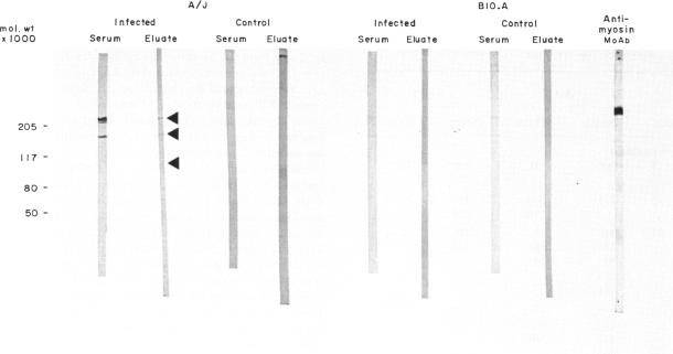
This study was undertaken to determine if immunoglobulin G (IgG) antibodies could be eluted from the hearts of mice with Coxsackievirus B3-induced autoimmune myocarditis and to characterize the immunoreactivity of any elutable autoantibodies. Susceptible (A/J) and resistant (B10.A) mice were administered the virus or the control treatment and killed at various times after treatment. Acid eluates from pooled heart tissue from each treatment group and each time were tested for IgG reactivity with normal heart tissue by immunohistochemistry and with normal heart extracts by Western immunostaining. Eluates from infected A/J mice reacted strongly with syngeneic heart and modestly with syngeneic skeletal muscle tissue. Eluates from infected B10.A or control mice of either strain exhibited little reactivity with either tissue. Tissue reactivity was similar when allogeneic tissue was used as the substrate. Eluates from infected A/J mice recognized the heavy chain of cardiac myosin and several other cardiac antigens by Western immunostaining while eluates from the other treatment groups exhibited little or no reactivity with any normal heart constituents. These results indicate that in vivo IgG deposition occurs in the hearts of mice with post-infectious autoimmune myocarditis and that the specificity of these antibodies is similar to that reported for serum from animals with this disease. The mechanism(s) leading to myocardial IgG deposition and its possible role in pathogenesis remain to be elucidated.
本研究旨在确定免疫球蛋白G(IgG)抗体是否可从小鼠心脏中洗脱,这些小鼠患有柯萨奇病毒B3诱导的自身免疫性心肌炎,并对任何可洗脱的自身抗体的免疫反应性进行表征。对易感(A/J)和抗性(B10.A)小鼠进行病毒或对照处理,并在处理后的不同时间处死。对每个处理组和每个时间点的合并心脏组织的酸性洗脱液进行检测,通过免疫组织化学检测其与正常心脏组织的IgG反应性,并通过Western免疫染色检测其与正常心脏提取物的反应性。感染的A/J小鼠的洗脱液与同基因心脏强烈反应,与同基因骨骼肌组织反应较弱。感染的B10.A或任一品系的对照小鼠的洗脱液与两种组织的反应性均较弱。当使用异基因组织作为底物时,组织反应性相似。通过Western免疫染色,感染的A/J小鼠的洗脱液识别心肌肌球蛋白重链和其他几种心脏抗原,而其他处理组的洗脱液与任何正常心脏成分的反应性较弱或无反应性。这些结果表明,在感染后自身免疫性心肌炎小鼠的心脏中发生了体内IgG沉积,并且这些抗体的特异性与患有该疾病动物血清中报道的特异性相似。导致心肌IgG沉积的机制及其在发病机制中的可能作用仍有待阐明。