Department of Microbiology, Tokyo Medical College, 6-1-1 Shinjuku, Tokyo 160.
Plant Physiol. 1990 Mar;92(3):802-8. doi: 10.1104/pp.92.3.802.
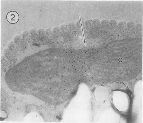
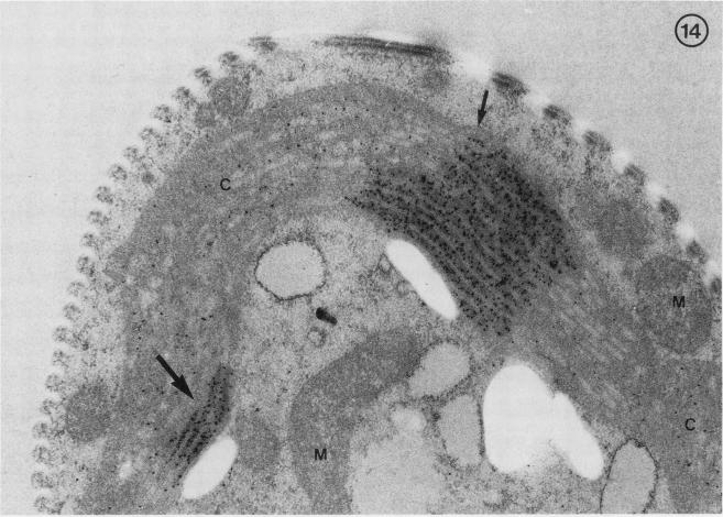
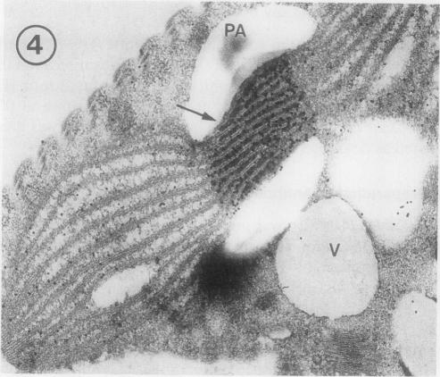
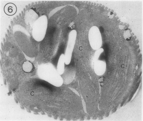
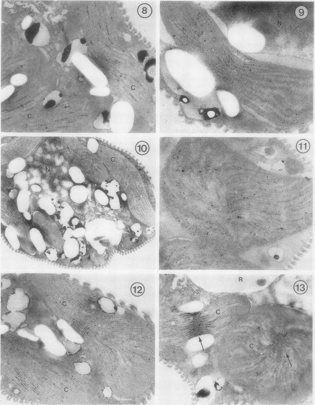
Euglena gracilis strain (Z) cells were synchronized under photoautotrophic conditions using a 14 hour light:10 hour dark regimen. The cells grew during the light period (growth phase) and divided during the following 10 hour period either in the dark or in the light (division phase). Changes in morphology of the pyrenoid and in the distribution of ribulose-1,5-bisphosphate carboxylase/oxygenase (Rubisco) within the chloroplasts were followed by immunoelectron microscopy during the growth and division phases of Euglena cells. Epon-embedded sections were labeled with an antibody to the holoenzyme followed by protein A-gold. The immunoreactive proteins were concentrated in the pyrenoid, and less densely distributed in the stroma during the growth phase. During the division phase, the pyrenoid could not be detected and the gold particles were dispersed throughout the stroma. Toward the end of the division phase, the pyrenoid began to form in the center of a chloroplast, and the immunoreactive proteins started to concentrate over that rudimentary pyrenoid. During the growth phase, small areas rich in gold particles, called ;satellite pyrenoid,' were observed, in addition to the main pyrenoid. From a comparison of photosynthetic CO(2)-fixation with the total carboxylase activity of Rubisco extracted from Euglena cells in the growth phase, it is suggested that the carboxylase in the pyrenoid functions in CO(2)-fixation in photosynthesis.
绿眼虫(Z)细胞在光自养条件下通过 14 小时光照:10 小时黑暗的周期进行同步化。细胞在光照期(生长阶段)生长,并在接下来的 10 小时内无论是在黑暗中还是在光照下(分裂阶段)进行分裂。在绿眼虫细胞的生长和分裂阶段,通过免疫电子显微镜观察淀粉核的形态变化和核酮糖-1,5-二磷酸羧化酶/加氧酶(Rubisco)在叶绿体中的分布。用针对全酶的抗体对包埋在环氧树脂中的切片进行标记,然后用蛋白 A-金进行标记。免疫反应蛋白集中在淀粉核中,在生长阶段在基质中分布较少。在分裂阶段,无法检测到淀粉核,金颗粒分散在基质中。在分裂阶段结束时,淀粉核开始在叶绿体的中心形成,免疫反应蛋白开始集中在那个原始淀粉核上。在生长阶段,除了主要淀粉核外,还观察到富含金颗粒的小区域,称为“卫星淀粉核”。通过比较光合作用中 CO2 的固定与从生长阶段的绿眼虫细胞中提取的 Rubisco 的总羧化酶活性,表明淀粉核中的羧化酶在光合作用中参与 CO2 的固定。