Pokharel Daya R, Rai Reeta, Kumar Pankaj, Chaturvedi C M, Rathaur Sushma
Department of Biochemistry, Faculty of Science, Banaras Hindu University, Varanasi-221005, India.
Filaria J. 2006 May 22;5:7. doi: 10.1186/1475-2883-5-7.
Like other helminth proteases, filarial proteases have also been shown to require for parasite survival inside the host and mediate various physiologic processes such as tissue invasion, feeding, embryogenesis and host immune evasion. Many of these proteases have shown potential for vaccines and chemotherapeutic agents against active filarial infections. Setaria cervi is a bovine filarial parasite and serves as a good parasite model for the studies in lymphatic filariasis. Recently, a 175 kDa collagenase and leucine aminopeptidase (LAP) have been purified and characterized from the bovine filarial parasite S. cervi and shown to be potential vaccine candidate and diagnostic marker, respectively for human lymphatic filariasis. However, their tissue localizations and putative roles in the parasite biology have not yet been examined and thus remain unclear. Therefore, the current study attempts to localize and explore the putative roles of these two enzymes in S. cervi.
The tissue distributions of 175 kDa collagenase and leucine aminopeptidase in S. cervi were examined by immunohistochemical and histochemical methods, respectively. Immune sera obtained from the jirds immunized with collagenase served as primary antibody, rabbit anti-mouse IgG-HRP conjugate as secondary antibody and DAB as the substrate for the immunostaining of collagenase. Leu-betaNA was used as the substrate for the histochemical staining of LAP.
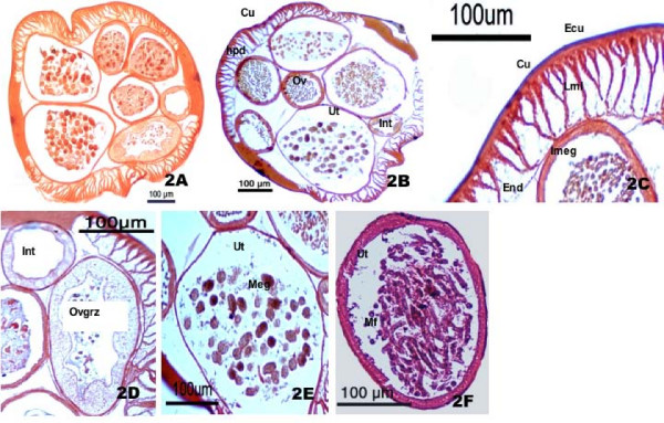
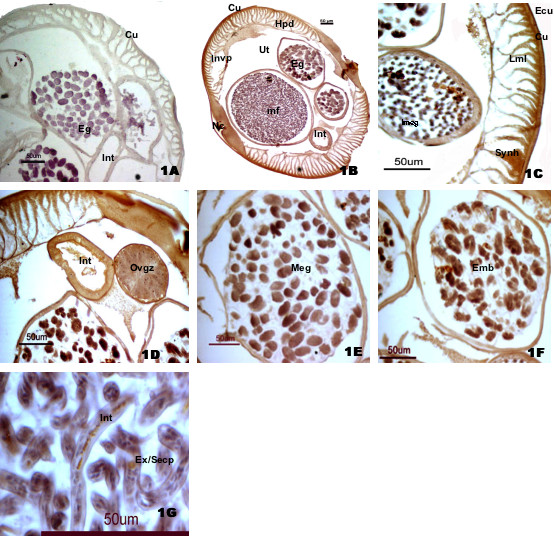
Both the collagenase and LAP were present in the body wall; however, they differ in their distribution pattern in different layers of body wall. Collagenase was mainly localized in epicuticle, cuticle, syncytial hypodermis and the nerve cord region whereas LAP was more concentrated in epicuticle, longitudinal muscle layers and almost absent or very faintly stained in syncytial hypodermis and nerve cord region. Both collagenase and LAP showed their common distributions in intestine, uterus and mature eggs, growing embryos and mf. Very strong immunostaining of collagenase in the outer body surface of the parasite indicates its major role in host-parasite relationship whereas the presence of LAP in muscular region suggests its role in tissue remodeling. The common presences of collagenase and LAP in the S. cervi intestine, ovary, uterus, eggs and mf suggest that they also have collaborative roles in molting, nutrition and embryogenesis. The data obtained on their immunological characterizations and their presence in important parasite organs give strong indication that they are critical for the survival of filarial parasite and thus can be good vaccine candidates and/or diagnostic markers for human lymphatic filariasis.
The manuscript reports for the first time the tissue distribution of collagenase and LAP in the bovine filarial parasite S. cervi and discuss their putative roles in vivo. Our findings also open the avenue to examine the roles of these two proteases in vivo, which will require further experiments like using their natural substrates and/or specific inhibitors in each tissues.
与其他蠕虫蛋白酶一样,丝虫蛋白酶也已被证明对寄生虫在宿主体内的存活至关重要,并介导各种生理过程,如组织侵袭、摄食、胚胎发生和宿主免疫逃避。这些蛋白酶中的许多已显示出作为抗活动性丝虫感染疫苗和化疗药物的潜力。鹿丝状线虫是一种牛丝虫寄生虫,是研究淋巴丝虫病的良好寄生虫模型。最近,已从牛丝虫寄生虫鹿丝状线虫中纯化并鉴定出一种175 kDa的胶原酶和亮氨酸氨肽酶(LAP),分别显示它们是人类淋巴丝虫病的潜在疫苗候选物和诊断标志物。然而,它们在组织中的定位以及在寄生虫生物学中的假定作用尚未得到研究,因此仍不清楚。因此,本研究试图定位并探索这两种酶在鹿丝状线虫中的假定作用。
分别采用免疫组织化学和组织化学方法检测鹿丝状线虫中175 kDa胶原酶和亮氨酸氨肽酶的组织分布。用胶原酶免疫沙鼠获得的免疫血清作为一抗,兔抗小鼠IgG-HRP共轭物作为二抗,DAB作为胶原酶免疫染色的底物。Leu-βNA用作LAP组织化学染色的底物。
胶原酶和LAP均存在于体壁中;然而,它们在体壁不同层中的分布模式不同。胶原酶主要定位于表皮、角质层、合胞体皮下组织和神经索区域,而LAP更集中于表皮、纵肌层,在合胞体皮下组织和神经索区域几乎不存在或染色非常浅。胶原酶和LAP在肠、子宫、成熟卵、发育中的胚胎和微丝蚴中均有共同分布。寄生虫体表外表面胶原酶的非常强的免疫染色表明其在宿主-寄生虫关系中的主要作用,而LAP在肌肉区域的存在表明其在组织重塑中的作用。胶原酶和LAP在鹿丝状线虫的肠、卵巢、子宫、卵和微丝蚴中的共同存在表明它们在蜕皮、营养和胚胎发生中也具有协同作用。关于它们免疫特性的数据以及它们在重要寄生虫器官中的存在有力地表明它们对丝虫寄生虫的存活至关重要,因此可以作为人类淋巴丝虫病的良好疫苗候选物和/或诊断标志物。
本手稿首次报道了胶原酶和LAP在牛丝虫寄生虫鹿丝状线虫中的组织分布,并讨论了它们在体内的假定作用。我们的发现也为研究这两种蛋白酶在体内的作用开辟了道路,这将需要进一步的实验,如在每个组织中使用它们的天然底物和/或特异性抑制剂。