Sutton Dwayne J, Tchounwou Paul B
Molecular Toxicology Research laboratory, NIH-Center for Environmental Health, College of Science, Engineering and Technology, Jackson State University, 1400 Lynch Street, Box 18540 Jackson, Mississippi 39217, USA.
Int J Environ Res Public Health. 2006 Mar;3(1):38-42. doi: 10.3390/ijerph2006030005.
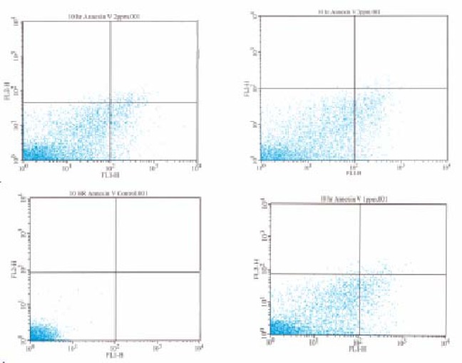
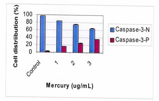
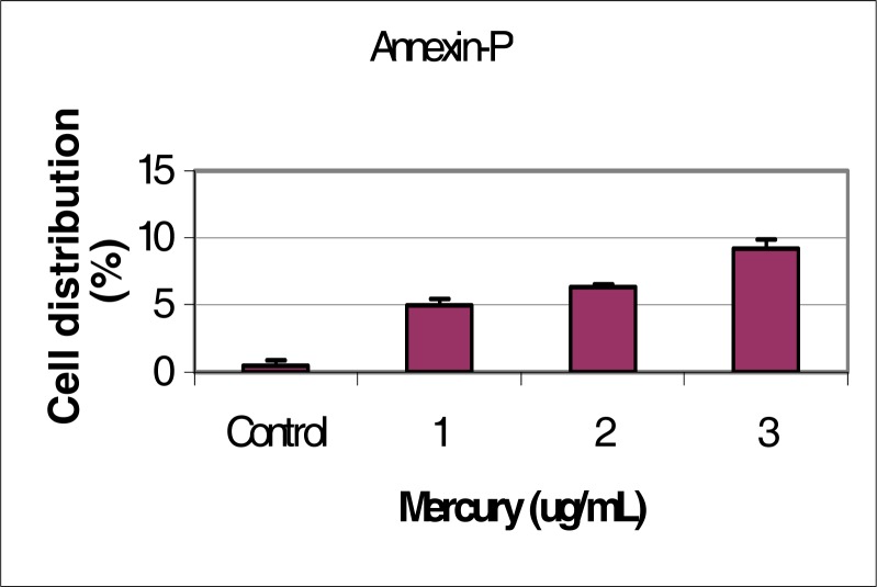
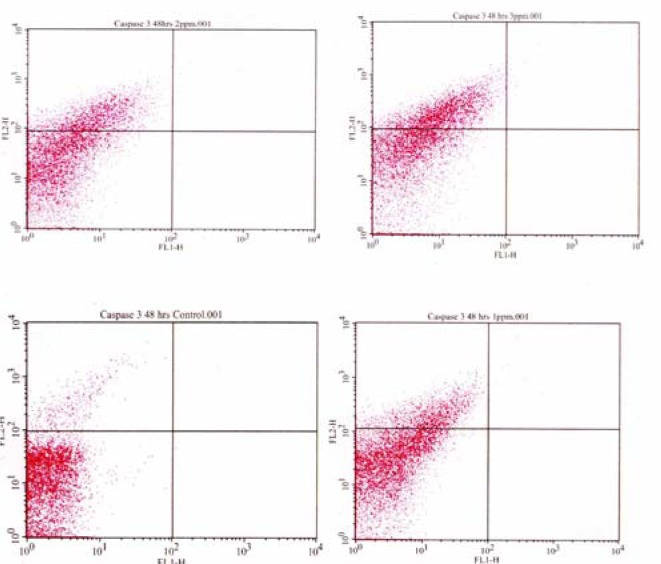
Apoptosis arises from the active initiation and propagation of a series of highly orchestrated specific biochemical events leading to the demise of the cell. It is a normal physiological process, which occurs during embryonic development as well as in the maintenance of tissue homeostasis. Diverse groups of molecules are involved in the apoptosis pathway and it functions as a mechanism to eliminate unwanted or irreparably damaged cells. However, inappropriate induction of apoptosis by environmental agents has broad ranging pathologic implications and has been associated with several diseases including cancer. The toxicity of several heavy metals such as mercury has been attributed to their high affinity to sulfhydryl groups of proteins and enzymes, and their ability to disrupt cell cycle progression and/or apoptosis in various tissues. The aim of this study was to assess the potential for mercury to induce early and late-stage apoptosis in human liver carcinoma (HepG2) cells. The Annexin-V and Caspase 3 assays were performed by flow cytometric analysis to determine the extent of phosphatidylserine externalization and Caspase 3 activation in mercury-treated HepG2 cells. Cells were exposed to mercury for 10 and 48 hours respectively at doses of 0, 1, 2, and 3 microg/mL based on previous cytotoxicity results in our laboratory indicating an LD50 of 3.5 +/- 0.6 microg/mL for mercury in HepG2 cells. The study data indicated a dose response relationship between mercury exposure and the degree of early and late-stage apoptosis in HepG2 cells. The percentages of cells undergoing early apoptosis were 0.03 +/- 0.03%, 5.19 +/- 0.04%, 6.36 +/- 0.04%, and 8.84 +/- 0.02% for 0, 1, 2, and 3 microg/mL of mercury respectively, indicating a gradual increase in apoptotic cells with increasing doses of mercury. The percentages of Caspase 3 positive cells undergoing late apoptosis were 3.58 +/- 0.03%, 17.06 +/- 0.05%, 23.32 +/- 0.03%, and 34.51 +/- 0.01% for 0, 1, 2, and 3 microg/mL of mercury respectively, also indicating a gradual increase in Caspase positive cells with increasing doses of mercury.
细胞凋亡源于一系列高度协调的特定生化事件的主动启动和传播,最终导致细胞死亡。它是一个正常的生理过程,发生在胚胎发育以及组织稳态的维持过程中。多种分子参与细胞凋亡途径,其作用是清除不需要的或无法修复的受损细胞。然而,环境因素不当诱导细胞凋亡具有广泛的病理意义,并与包括癌症在内的多种疾病相关。几种重金属(如汞)的毒性归因于它们对蛋白质和酶的巯基具有高亲和力,以及它们破坏各种组织中细胞周期进程和/或细胞凋亡的能力。本研究的目的是评估汞诱导人肝癌(HepG2)细胞早期和晚期凋亡的潜力。通过流式细胞术分析进行膜联蛋白V和半胱天冬酶3检测,以确定汞处理的HepG2细胞中磷脂酰丝氨酸外化程度和半胱天冬酶3激活程度。根据我们实验室先前的细胞毒性结果,细胞分别以0、1、2和3微克/毫升的剂量暴露于汞中10小时和48小时,结果表明汞对HepG2细胞的半数致死剂量为3.5±0.6微克/毫升。研究数据表明,汞暴露与HepG2细胞早期和晚期凋亡程度之间存在剂量反应关系。汞浓度为0、1、2和3微克/毫升时,早期凋亡细胞的百分比分别为0.03±0.03%、5.19±0.04%、6.36±0.04%和8.84±0.02%,表明随着汞剂量增加,凋亡细胞逐渐增多。汞浓度为0、1、2和3微克/毫升时,晚期凋亡的半胱天冬酶3阳性细胞的百分比分别为3.58±0.03%、17.06±0.05%、23.32±0.03%和34.51±0.01%,也表明随着汞剂量增加,半胱天冬酶阳性细胞逐渐增多。