Department of Pharmacy, Henan University of Science and Technology, 263 Kaiyuan Avenue, Luoyang, 471023, China.
J Exp Clin Cancer Res. 2018 Jul 9;37(1):142. doi: 10.1186/s13046-018-0823-2.
JS-K is a nitric oxide (NO) donor and could generate intracellularly high levels of NO. The study explores PP2A as a tumor suppressor is a major determinant mediating JS-K-caused apoptosis in human hepatocellular carcinoma (HCC) cells.
The human HCC cell lines (PLC5, Huh-7, Bel-7402, SMMC-7721 and HepG2) were used to assess effects of JS-K on cell viability, apoptosis induction and PP2A activation. Effects of JS-K on cell morphology, mitochondrial membrane potential, apoptosis and NO levels were determined in HCC cells expressing PP2A. Simultaneously, the expression of PP2A family including PP2A-A(α/β), PP2A-B55, PP2A-C(α/β) and the substrates of PP2A, such as β-catenin, c-Myc and p-Bcl-2 (Ser70) were detected in sensitive HCC cells. Furthermore, the role of NO in mediating the expression of PP2A was further validated with Z-VAD-FMK (a caspase inhibitor), Carboxy-PTIO (a NO scavenger), okadaic acid (OA, a PP2A inhibitor) and FTY720 (a PP2A agonist) in JS-K treated cells. In addition, the genetic manuplation of PP2A including overexpression and knockdown have been also performed in JS-K treated cells. Moreover, the rat model of primary hepatic carcinoma was established with diethylnitrosamine for 16 weeks to verify the anti-tumor effects of JS-K in vivo. Immunohistochemical and Western blot analysis were used to determine the expression of proteins in rat primary hepatic carcinoma tissues.
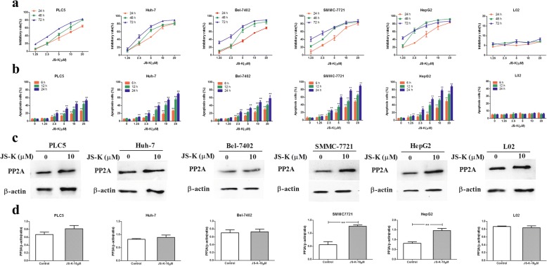
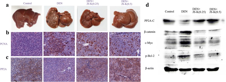
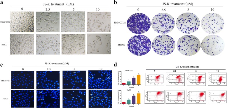
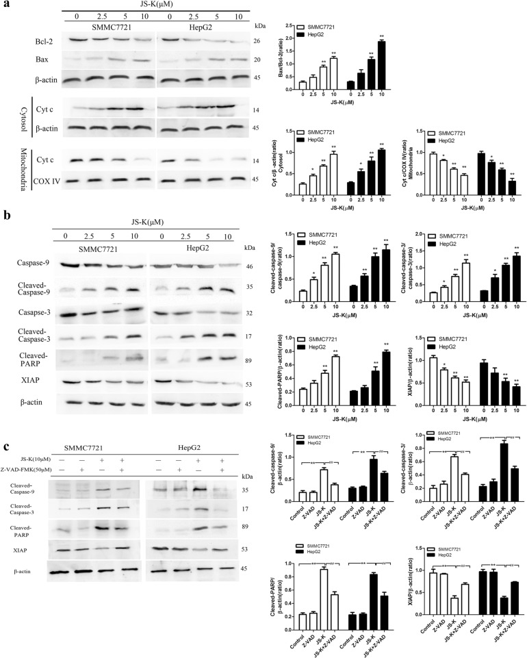
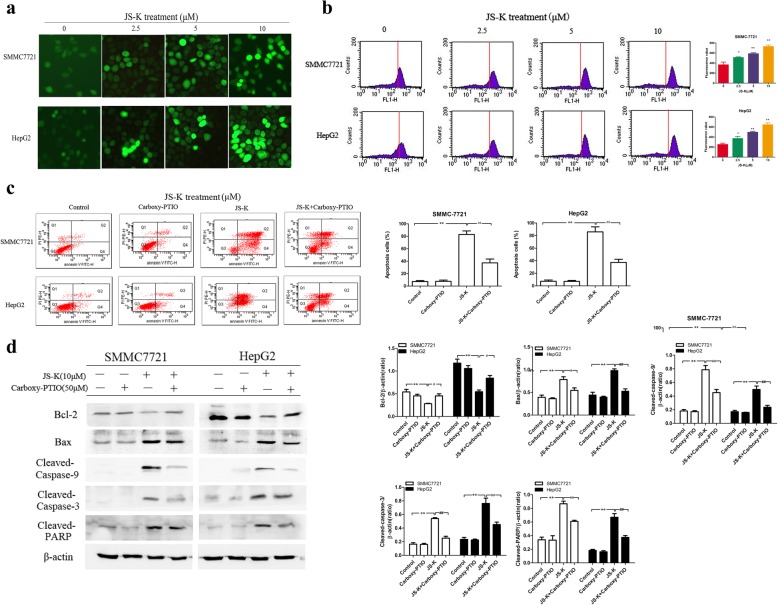
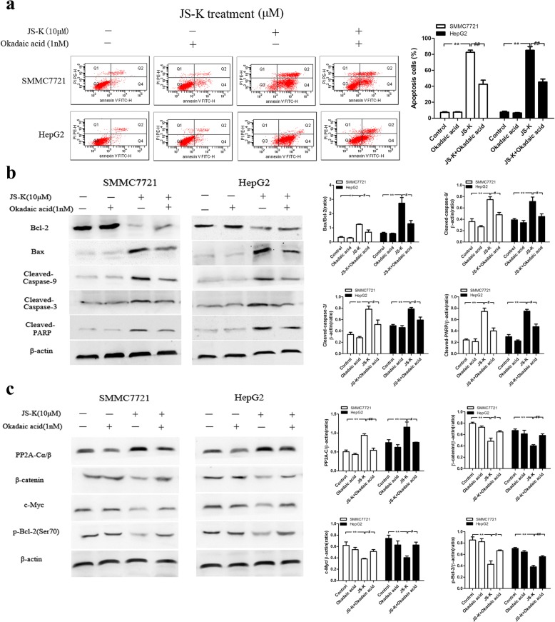
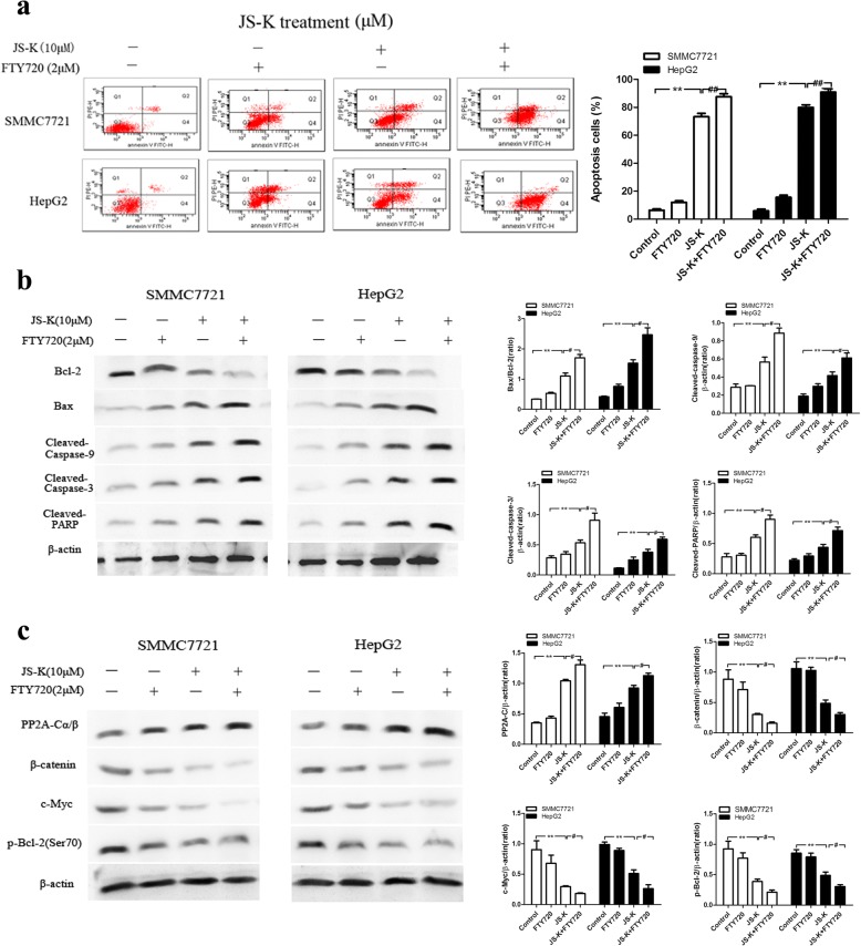
JS-K significantly inhibited cell proliferation, increased apoptosis rate and activated PP2A activity in five HCC cells viability, especially SMMC7721 and HepG2 cells. It was characterized by loss of mitochondrial membrane potential, significant externalization of phosphatidylserine, nuclear morphological changes. Moreover, JS-K enhanced Bax-to-Bcl-2 ratio, released cytochrome c (Cyt c) from mitochondria, activated cleaved-caspase-9/3 and the cleavage of PARP, and decreased the expression of X-linked inhibitor of apoptosis protein (XIAP). Both Z-VAD-FMK and Carboxy-PTIO suppressed the activation of cleaved-caspase-9/3 and of cleaved-PARP in JS-K-treated sensitive HCC cells. Simultaneously, JS-K treatment could lead to the activation of protein phosphatase 2A-C (PP2A-C) but not PP2A-A and PP2A-B55, which subsequently inactivated and dephosphorylated the PP2A substrates including β-catenin, c-Myc, and p-Bcl-2 (Ser70). However, silencing PP2A-C could abolish both the activation of PP2A-C and down-regulation of β-catenin, c-Myc and p-Bcl-2 (Ser70) in sensitive HCC cells. Conversely, PP2A overexpression could enhance the effects of JS-K on activation of PP2A and down-regulation of β-catenin, c-Myc and p-Bcl-2 (Ser70). In addition, adding okadaic acid (OA), a PP2A inhibitor, abolished the effects of JS-K on apoptosis induction, PP2A activation and the substrates of PP2A dephosphorylation; FTY720, a PP2A agonist, enhanced the effects of JS-K including apoptosis induction, PP2A activation and the substrates of PP2A dephosphorylation. The mice exhibited a lower number and smaller tumor nodules in response to JS-K-treated group. A marked increase in the number of hepatocytes with PCNA-positive nuclei (proliferating cells) was evident in DEN group and tended to decrease with JS-K treatment. Furthermore, JS-K treatment could induce PP2A activation and the substrates of PP2A inactivation such as β-catenin, c-Myc and p-Bcl-2(Ser70) in DEN-induced hepatocarcinogenesis.
High levels of NO released from JS-K induces a caspase-dependent apoptosis through PP2A activation.
JS-K 是一种一氧化氮(NO)供体,可以在细胞内产生高水平的 NO。本研究探讨了蛋白磷酸酶 2A(PP2A)作为一种肿瘤抑制因子,是介导 JS-K 诱导人肝癌(HCC)细胞凋亡的主要决定因素。
使用人 HCC 细胞系(PLC5、Huh-7、Bel-7402、SMMC-7721 和 HepG2)评估 JS-K 对细胞活力、细胞凋亡诱导和 PP2A 激活的影响。在表达 PP2A 的 HCC 细胞中,测定 JS-K 对细胞形态、线粒体膜电位、细胞凋亡和 NO 水平的影响。同时,检测敏感 HCC 细胞中 PP2A 家族包括 PP2A-A(α/β)、PP2A-B55、PP2A-C(α/β)以及 PP2A 的底物,如β-连环蛋白、c-Myc 和 p-Bcl-2(Ser70)的表达。此外,使用 Z-VAD-FMK(一种半胱天冬酶抑制剂)、Carboxy-PTIO(一种 NO 清除剂)、OA(一种 PP2A 抑制剂)和 FTY720(一种 PP2A 激动剂)进一步验证 NO 在介导 PP2A 表达中的作用在 JS-K 处理的细胞中。此外,还在 JS-K 处理的细胞中进行了包括过表达和敲低在内的 PP2A 的遗传操作。此外,使用二乙基亚硝胺(DEN)建立大鼠原发性肝癌模型,以验证 JS-K 在体内的抗肿瘤作用。免疫组织化学和 Western blot 分析用于确定大鼠原发性肝癌组织中蛋白质的表达。
JS-K 显著抑制了 5 种 HCC 细胞的增殖,增加了细胞凋亡率,激活了细胞的 PP2A 活性,特别是 SMMC7721 和 HepG2 细胞。其特征是线粒体膜电位丧失,磷脂酰丝氨酸显著外翻,核形态变化。此外,JS-K 增强了 Bax-to-Bcl-2 比值,从线粒体中释放细胞色素 c(Cyt c),激活 cleaved-caspase-9/3 和 PARP 的裂解,并降低了 X 连锁凋亡抑制蛋白(XIAP)的表达。Z-VAD-FMK 和 Carboxy-PTIO 均抑制了 JS-K 处理的敏感 HCC 细胞中 cleaved-caspase-9/3 和 cleaved-PARP 的激活。同时,JS-K 处理可导致蛋白磷酸酶 2A-C(PP2A-C)的激活,但不激活 PP2A-A 和 PP2A-B55,随后使 PP2A 的底物包括β-连环蛋白、c-Myc 和 p-Bcl-2(Ser70)失活和去磷酸化。然而,沉默 PP2A-C 可消除敏感 HCC 细胞中 PP2A-C 的激活和β-连环蛋白、c-Myc 和 p-Bcl-2(Ser70)的下调。相反,PP2A 的过表达可以增强 JS-K 对 PP2A 的激活和β-连环蛋白、c-Myc 和 p-Bcl-2(Ser70)的下调作用。此外,加入 OA(一种 PP2A 抑制剂)可消除 JS-K 对细胞凋亡诱导、PP2A 激活和 PP2A 底物去磷酸化的作用;FTY720(一种 PP2A 激动剂)增强了 JS-K 的作用,包括细胞凋亡诱导、PP2A 激活和 PP2A 底物的去磷酸化。小鼠对 JS-K 处理组的肿瘤数量和肿瘤结节大小均较低。DEN 组的肝细胞 PCNA 阳性核(增殖细胞)数量明显增加,且随着 JS-K 处理而趋于减少。此外,JS-K 处理可诱导 DEN 诱导的肝癌发生中 PP2A 的激活和 PP2A 底物如β-连环蛋白、c-Myc 和 p-Bcl-2(Ser70)的失活。
JS-K 释放的高水平 NO 通过激活 PP2A 诱导 caspase 依赖性细胞凋亡。