Hasan Zahra, Ashraf Mussarat, Tayyebi Ali, Hussain Rabia
Department of Pathology and Microbiology, The Aga Khan University, Karachi, Pakistan.
BMC Microbiol. 2006 Sep 18;6:78. doi: 10.1186/1471-2180-6-78.
Virulent Mycobacterium leprae interfere with host defense mechanisms such as cytokine activation and apoptosis. The mitochondrial pathway of apoptosis is regulated by the Bcl-2 family of proteins. Expression of Fas ligand and apoptotic proteins is found in leprosy lesions and M. leprae has been shown to activate pro-apoptotic Bcl-2 genes, Bak and Bax. However, the mechanism by which M. leprae modulates apoptosis is as yet unclear. We investigated expression of apoptotic genes in THP-1 monocytes in response to infection by M. leprae and non-pathogenic M. bovis BCG.
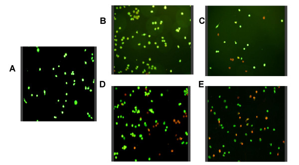
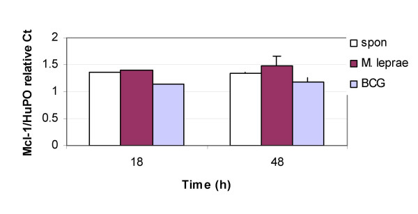
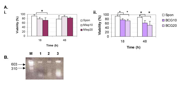
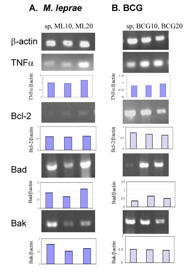
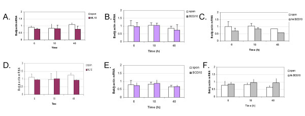
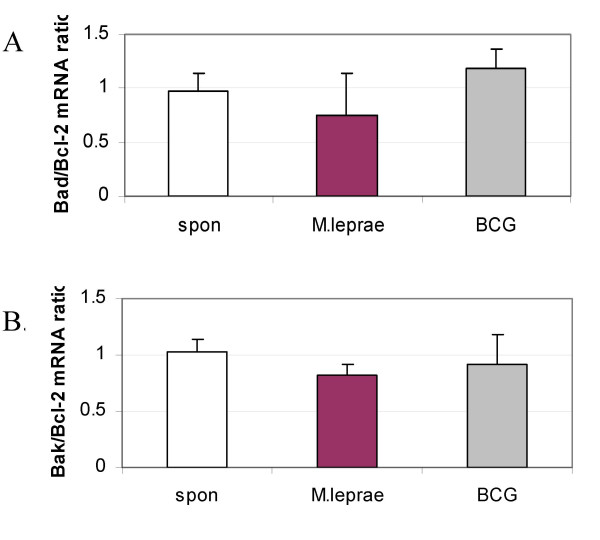
M. leprae did not induce apoptosis in THP-1 cells, while BCG induced a significant loss of cell viability by 18 h post-infection at both (multiplicity of infection) MOI-10 and 20, with an increase by 48 h. BCG-induced cell death was accompanied by characteristic apoptotic DNA laddering in cells. Non-viable BCG had a limited effect on host cell death suggesting that BCG-induced apoptosis was a function of mycobacterial viability. M. leprae also activated lower levels of TNF-alpha secretion and TNF-alpha mRNA expression than BCG. Mycobacterium-induced activation of apoptotic gene expression was determined over a time course of infection. M. leprae reduced Bad and Bak mRNA expression by 18 h post-stimulation, with a further decrease at 48 h. Outcome of cell viability is determined by the ratio between pro- and anti-apoptotic proteins present in the cell. M. leprae infection resulted in downregulation of gene expression ratios, Bad/Bcl-2 mRNA by 39% and Bak/Bcl-2 mRNA by 23%. In contrast, live BCG increased Bad/Bcl-2 mRNA (29 %) but had a negligible effect on Bak/Bcl-2 mRNA. Heat killed BCG induced only a negligible (1-4 %) change in mRNA expression of either Bak/Bcl-2 or Bad/Bcl-2. Additionally, M. leprae upregulated the expression of anti-apoptotic gene Mcl-1 while, BCG downregulated Mcl-1 mRNA.
This study proposes an association between mycobacterium-induced apoptosis in THP-1 cells and the regulation of Bcl-2 family of proteins. M. leprae restricts apoptosis in THP-1 cells by downregulation of Bad and Bak and upregulation of Mcl-1 mRNA expression.
强毒力麻风分枝杆菌会干扰宿主防御机制,如细胞因子激活和细胞凋亡。细胞凋亡的线粒体途径受Bcl-2蛋白家族调控。在麻风病皮损中发现了Fas配体和凋亡蛋白的表达,并且已证明麻风分枝杆菌可激活促凋亡Bcl-2基因Bak和Bax。然而,麻风分枝杆菌调节细胞凋亡的机制尚不清楚。我们研究了麻风分枝杆菌和非致病性牛分枝杆菌卡介苗(BCG)感染后THP-1单核细胞中凋亡基因的表达情况。
麻风分枝杆菌未诱导THP-1细胞凋亡,而BCG在感染复数(MOI)为10和20时,在感染后18小时诱导细胞活力显著丧失,48小时时有所增加。BCG诱导的细胞死亡伴随着细胞中典型的凋亡DNA梯状条带。无活力的BCG对宿主细胞死亡的影响有限,这表明BCG诱导的细胞凋亡是分枝杆菌活力的一种功能。与BCG相比,麻风分枝杆菌激活的肿瘤坏死因子-α(TNF-α)分泌水平和TNF-α mRNA表达水平也较低。在感染的时间进程中测定了分枝杆菌诱导的凋亡基因表达激活情况。麻风分枝杆菌在刺激后18小时降低了Bad和Bak mRNA表达,48小时时进一步降低。细胞活力的结果取决于细胞中促凋亡蛋白和抗凋亡蛋白之间的比例。麻风分枝杆菌感染导致基因表达比例下调,Bad/Bcl-2 mRNA下调39%,Bak/Bcl-2 mRNA下调23%。相比之下,活BCG使Bad/Bcl-2 mRNA增加(29%),但对Bak/Bcl-2 mRNA的影响可忽略不计。热灭活的BCG对Bak/Bcl-2或Bad/Bcl-2的mRNA表达仅诱导可忽略不计的(1 - 4%)变化。此外,麻风分枝杆菌上调了抗凋亡基因Mcl-1的表达,而BCG下调了Mcl-1 mRNA。
本研究提出了分枝杆菌诱导THP-1细胞凋亡与Bcl-2蛋白家族调控之间的关联。麻风分枝杆菌通过下调Bad和Bak以及上调Mcl-1 mRNA表达来限制THP-1细胞凋亡。