Wood Bayden R, Bambery Keith R, Evans Corey J, Quinn Michael A, McNaughton Don
Centre for Biospectroscopy and School of Chemistry, Monash University, 3800 Victoria, Australia.
BMC Med Imaging. 2006 Oct 3;6:12. doi: 10.1186/1471-2342-6-12.
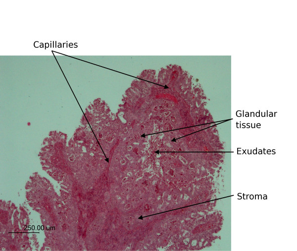
Three-dimensional (3D) multivariate Fourier Transform Infrared (FTIR) image maps of tissue sections are presented. A villoglandular adenocarcinoma from a cervical biopsy with a number of interesting anatomical features was used as a model system to demonstrate the efficacy of the technique.
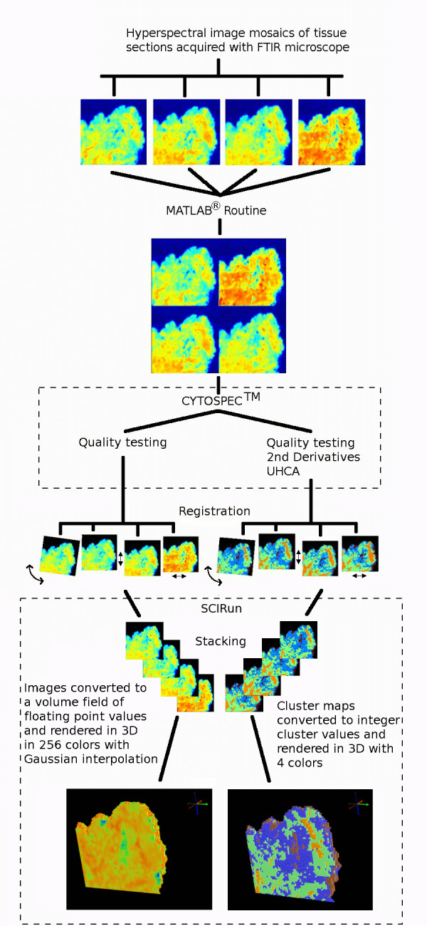
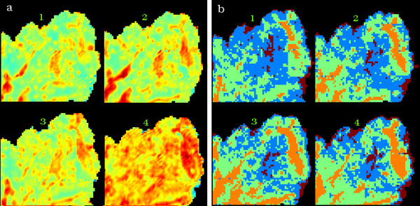
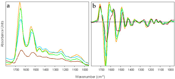
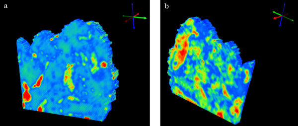
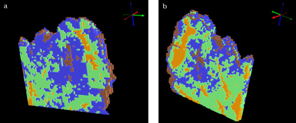
Four FTIR images recorded using a focal plane array detector of adjacent tissue sections were stitched together using a MATLAB routine and placed in a single data matrix for multivariate analysis using Cytospec. Unsupervised Hierarchical Cluster Analysis (UHCA) was performed simultaneously on all 4 sections and 4 clusters plotted. The four UHCA maps were then stacked together and interpolated with a box function using SCIRun software.
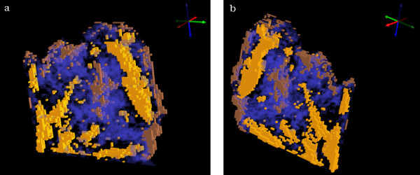
The resultant 3D-images can be rotated in three-dimensions, sliced and made semi-transparent to view the internal structure of the tissue block. A number of anatomical and histopathological features including connective tissue, red blood cells, inflammatory exudate and glandular cells could be identified in the cluster maps and correlated with Hematoxylin & Eosin stained sections. The mean extracted spectra from individual clusters provide macromolecular information on tissue components.
3D-multivariate imaging provides a new avenue to study the shape and penetration of important anatomical and histopathological features based on the underlying macromolecular chemistry and therefore has clear potential in biology and medicine.
展示了组织切片的三维(3D)多变量傅里叶变换红外(FTIR)图像图谱。来自宫颈活检的具有一些有趣解剖特征的绒毛腺腺癌被用作模型系统来证明该技术的有效性。
使用MATLAB程序将使用焦平面阵列探测器记录的相邻组织切片的四张FTIR图像拼接在一起,并放置在单个数据矩阵中,使用Cytospec进行多变量分析。对所有4个切片同时进行无监督层次聚类分析(UHCA)并绘制4个聚类图。然后将这四个UHCA图谱堆叠在一起,并使用SCIRun软件用盒式函数进行插值。
所得的3D图像可以在三维空间中旋转、切片并设置为半透明以查看组织块的内部结构。在聚类图中可以识别出包括结缔组织、红细胞、炎性渗出物和腺细胞在内的许多解剖和组织病理学特征,并与苏木精和伊红染色切片相关联。从各个聚类中提取的平均光谱提供了关于组织成分的大分子信息。
3D多变量成像基于潜在的大分子化学提供了一条研究重要解剖和组织病理学特征的形状和穿透性的新途径,因此在生物学和医学中具有明显的潜力。